
Page 289 - Read Online
P. 289
Page 4 of 8 Goh et al. Mini-invasive Surg 2021;5:30 https://dx.doi.org/10.20517/2574-1225.2021.42
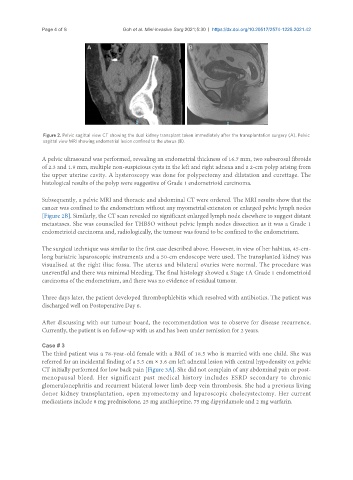
Figure 2. Pelvic sagittal view CT showing the dual kidney transplant taken immediately after the transplantation surgery (A). Pelvic
sagittal view MRI showing endometrial lesion confined to the uterus (B).
A pelvic ultrasound was performed, revealing an endometrial thickness of 16.7 mm, two subserosal fibroids
of 2.3 and 1.9 mm, multiple non-suspicious cysts in the left and right adnexa and a 2-cm polyp arising from
the upper uterine cavity. A hysteroscopy was done for polypectomy and dilatation and curettage. The
histological results of the polyp were suggestive of Grade 1 endometrioid carcinoma.
Subsequently, a pelvic MRI and thoracic and abdominal CT were ordered. The MRI results show that the
cancer was confined to the endometrium without any myometrial extension or enlarged pelvic lymph nodes
[Figure 2B]. Similarly, the CT scan revealed no significant enlarged lymph node elsewhere to suggest distant
metastases. She was counselled for THBSO without pelvic lymph nodes dissection as it was a Grade 1
endometrioid carcinoma and, radiologically, the tumour was found to be confined to the endometrium.
The surgical technique was similar to the first case described above. However, in view of her habitus, 45-cm-
long bariatric laparoscopic instruments and a 50-cm endoscope were used. The transplanted kidney was
visualised at the right iliac fossa. The uterus and bilateral ovaries were normal. The procedure was
uneventful and there was minimal bleeding. The final histology showed a Stage 1A Grade 1 endometrioid
carcinoma of the endometrium, and there was no evidence of residual tumour.
Three days later, the patient developed thrombophlebitis which resolved with antibiotics. The patient was
discharged well on Postoperative Day 6.
After discussing with our tumour board, the recommendation was to observe for disease recurrence.
Currently, the patient is on follow-up with us and has been under remission for 2 years.
Case # 3
The third patient was a 78-year-old female with a BMI of 18.5 who is married with one child. She was
referred for an incidental finding of a 5.5 cm × 3.6 cm left adnexal lesion with central hypodensity on pelvic
CT initially performed for low back pain [Figure 3A]. She did not complain of any abdominal pain or post-
menopausal bleed. Her significant past medical history includes ESRD secondary to chronic
glomerulonephritis and recurrent bilateral lower limb deep vein thrombosis. She had a previous living
donor kidney transplantation, open myomectomy and laparoscopic cholecystectomy. Her current
medications include 8 mg prednisolone, 25 mg azathioprine, 75 mg dipyridamole and 2 mg warfarin.