
Page 288 - Read Online
P. 288
Goh et al. Mini-invasive Surg 2021;5:30 https://dx.doi.org/10.20517/2574-1225.2021.42 Page 3 of 8
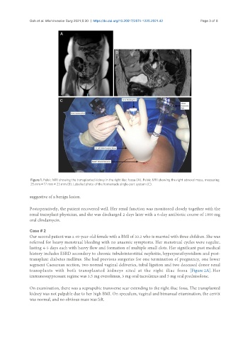
Figure 1. Pelvic MRI showing the transplanted kidney in the right iliac fossa (A). Pelvic MRI showing the right adnexal mass, measuring
25 mm × 17 mm × 23 mm (B). Labelled photo of the homemade single-port system (C).
suggestive of a benign lesion.
Postoperatively, the patient recovered well. Her renal function was monitored closely together with the
renal transplant physician, and she was discharged 2 days later with a 6-day antibiotic course of 1800 mg
oral clindamycin.
Case # 2
Our second patient was a 49-year-old female with a BMI of 33.2 who is married with three children. She was
referred for heavy menstrual bleeding with no anaemic symptoms. Her menstrual cycles were regular,
lasting 4-5 days each with heavy flow and formation of multiple small clots. Her significant past medical
history includes ESRD secondary to chronic tubulointerstitial nephritis, hyperparathyroidism and post-
transplant diabetes mellitus. She had previous surgeries for one termination of pregnancy, one lower
segment Caesarean section, two normal vaginal deliveries, tubal ligation and two deceased donor renal
transplants with both transplanted kidneys sited at the right iliac fossa [Figure 2A]. Her
immunosuppressant regime was 3.5 mg everolimus, 3 mg oral tacrolimus and 5 mg oral prednisolone.
On examination, there was a suprapubic transverse scar extending to the right iliac fossa. The transplanted
kidney was not palpable due to her high BMI. On speculum, vaginal and bimanual examination, the cervix
was normal, and no obvious mass was felt.