
Page 923 - Read Online
P. 923
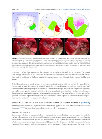
Page 6 of 11                                         Azab et al. Mini-invasive Surg 2020;4:88  I  http://dx.doi.org/10.20517/2574-1225.2020.75


               Figure 5. Endoscope-controlled supraorbital keyhole eyebrow approach. A: head position, skin incision, burr hole placement and
               craniotomy design; B: initial endoscopic view gained through right-sided approach; C: further brain relaxation and panoramic exposure
               of a tuberculum sellae meningioma; D-F: intraoperative endoscopic views of tuberculum sellae meningioma (asterisk) being exposed
               with plane development and bipolar coagulation, left-sided approach. A1: first segment of anterior cerebral artery; ACP: anterior clinoid
               process; ICA: internal carotid artery; FL: frontal lobe; OC: optic chiasm; ON: optic nerve; PS: planum sphenoidale; TS: tuberculum sellae
               (Illustrations A through C by Waleed Azab)


               consequence of the light source and the viewing lens being located outside the craniotomy. The loss of
               light energy at the edges of the small craniotomy and the dropped shadows on the structures within the
               field further contribute to the lesser quality of the microscopic view obtained during supraorbital keyhole
               surgery.


               Notwithstanding, some disadvantages of endoscope-assisted surgery exist and include the lack of three-
               dimensionality, need for familiarity with endoscopic devices, need to develop eye-hand coordination, and
                                                        [34]
               imitation of the operating range of instruments . Such disadvantages, however, are largely outweighed by
               the higher visual quality, surgical radicality and lesser complication profile offered by this type of surgery.
               In our opinion, rigid endoscopes are indispensable components of the array of surgical tools required to
               perform a keyhole supraorbital approach. We truly believe endoscopes will completely replace surgical
               microscopes for this type of surgery in the future.


               SURGICAL TECHNIQUE OF THE SUPRAORBITAL KEYHOLE EYEBROW APPROACH [FIGURE 5]
               The surgical technique of the supraorbital keyhole eyebrow approach has been extensively described in the
               literature [19,29,35,41-46] . A brief description of the technique will be given below.


               Preoperative planning
               Careful case selection is paramount when operating via the supraorbital eyebrow approach. The patient’s
               individual anatomy should be thoroughly evaluated. One important consideration is the lateral extent
               of the frontal air sinus which dictates the medial border of the supraorbital craniotomy and determines
               whether an appropriate surgical trajectory would be possible. Meningiomas with high superior extent need
               more retroflexion of the head to obtain a proper working trajectory. In general, the closer the tumor to the