
Page 921 - Read Online
P. 921
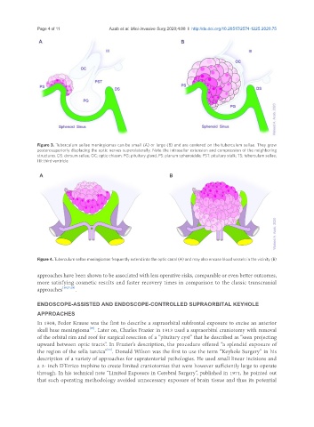
Page 4 of 11                                         Azab et al. Mini-invasive Surg 2020;4:88  I  http://dx.doi.org/10.20517/2574-1225.2020.75


               Figure 3. Tuberculum sellae meningiomas can be small (A) or large (B) and are centered on the tuberculum sellae. They grow
               posterosuperiorly displacing the optic nerves superolaterally. Note the intrasellar extension and compression of the neighboring
               structures. DS: dorsum sellae; OC: optic chiasm; PG: pituitary gland; PS: planum sphenoidale; PST: pituitary stalk; TS: tuberculum sellae;
               III: third ventricle


               Figure 4. Tuberculum sellae meningiomas frequently extend into the optic canal (A) and may also encase blood vessels in the vicinity (B)

               approaches have been shown to be associated with less operative risks, comparable or even better outcomes,
               more satisfying cosmetic results and faster recovery times in comparison to the classic transcranial
               approaches [19,27,29] .


               ENDOSCOPE-ASSISTED AND ENDOSCOPE-CONTROLLED SUPRAORBITAL KEYHOLE
               APPROACHES
               In 1908, Fedor Krause was the first to describe a supraorbital subfrontal exposure to excise an anterior
                                   [30]
               skull base meningioma . Later on, Charles Frazier in 1913 used a supraorbital craniotomy with removal
               of the orbital rim and roof for surgical resection of a “pituitary cyst” that he described as “seen projecting
               upward between optic tracts”. In Frazier’s description, the procedure offered “a splendid exposure of
                                          [31]
               the region of the sella turcica” . Donald Wilson was the first to use the term “Keyhole Surgery” in his
               description of a variety of approaches for supratentorial pathologies. He used small linear incisions and
               a 2- inch D’Errico trephine to create limited craniotomies that were however sufficiently large to operate
               through. In his technical note “Limited Exposure in Cerebral Surgery”, published in 1971, he pointed out
               that such operating methodology avoided unnecessary exposure of brain tissue and thus its potential