
Page 920 - Read Online
P. 920
Azab et al. Mini-invasive Surg 2020;4:88 I http://dx.doi.org/10.20517/2574-1225.2020.75 Page 3 of 11
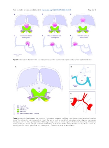
Figure 1. Nomenclature of anterior skull base meningiomas according to anatomical origin in axial (A-C) and sagittal (D-F) views
Figure 2. Anatomical environment and structures often related to anterior skull base meningioma. A: axial overview; B: sagittal
view of the sellar region and structures in its vicinity that may be involved especially in tuberculum sellae and planum sphenoidale
meningiomas; C: view of the optic apparatus and the neighboring vasculature. A1: first segment of anterior cerebral artery; ACP: anterior
clinoid process; DS: dorsum sellae; ICA: internal carotid artery; MCA: middle cerebral artery; OC: optic chiasm; ON: optic nerve; PG:
pituitary gland; PS: planum sphenoidale; PST: pituitary stalk; TS: tuberculum sellae; III: third ventricle