
Page 611 - Read Online
P. 611
De Iaco et al. Mini-invasive Surg 2020;4:63 I http://dx.doi.org/10.20517/2574-1225.2020.37 Page 5 of 16
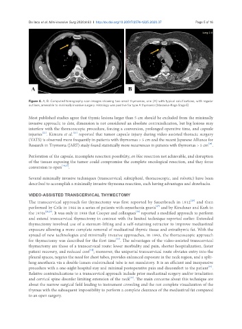
Figure 4. A, B: Computed tomography scan images showing two small thymomas, one (A) with typical calcifications, with regular
outlines, amenable to minimally invasive surgery. Histology was positive for type A thymoma (Masaoka-Koga Stage I)
Most published studies agree that thymic lesions larger than 5 cm should be excluded from the minimally
invasive approach; to date, dimension is not considered an absolute contraindication, but big lesions may
interfere with the thoracoscopic procedure, forcing a conversion, prolonged operative time, and capsule
[23]
[22]
injuries . Kimura et al. reported that tumor capsule injury during video-assisted thoracic surgery
(VATS) is observed more frequently in patients with thymomas > 5 cm and the recent Japanese Alliance for
[24]
Research in Thymoma (JART) study found statistically more recurrences in patients with thymomas > 5 cm .
Perforation of the capsule, incomplete resection possibility, en bloc resection not achievable, and disruption
of the tissues exposing the tumor could compromise the complete oncological resection, and they force
conversion to open [10,25] .
Several minimally invasive techniques (transcervical, subxiphoid, thoracoscopic, and robotic) have been
described to accomplish a minimally invasive thymoma resection, each having advantages and drawbacks.
VIDEO-ASSISTED TRANSCERVICAL THYMECTOMY
The transcervical approach for thymectomy was first reported by Sauerbruch in 1912 and then
[26]
[27]
performed by Crile in 1966 in a series of patients with myasthenia gravis and by Kirschner and Kark in
[30]
the 1970s [28,29] . It was only in 1988 that Cooper and colleagues reported a modified approach to perform
and extend transcervical thymectomy in contrast with the limited technique reported earlier. Extended
thymectomy involved use of a sternum-lifting and a self-retaining retractor to improve mediastinal
exposure allowing a more complete removal of mediastinal thymic tissue and extrathymic fat. With the
spread of new technologies and minimally invasive approaches, in 1993, the thoracoscopic approach
[31]
for thymectomy was described for the first time . The advantages of the video-assisted transcervical
thymectomy are those of a transcervical route: lower morbidity and pain, shorter hospitalization, faster
[32]
patient recovery, and reduced cost ; moreover, the uniportal transcervical route obviates entry into the
pleural spaces, negates the need for chest tubes, provides enhanced exposure in the neck region, and a split-
lung anesthesia via a double-lumen endotracheal tube is not mandatory. It is an efficient and inexpensive
[33]
procedure with a one-night hospital stay and minimal postoperative pain and discomfort to the patient .
Relative contraindications to a transcervical approach include prior mediastinal surgery and/or irradiation
and cervical spine disorder limiting extension of the neck . The main concerns about this technique are
[33]
about the narrow surgical field leading to instrument crowding and the not complete visualization of the
thymus with the subsequent impossibility to perform a complete clearance of the mediastinal fat compared
to an open surgery.