
Page 52 - Read Online
P. 52
Kawada et al. Mini-invasive Surg 2020;4:7 I http://dx.doi.org/10.20517/2574-1225.2019.44 Page 3 of 6
A
B
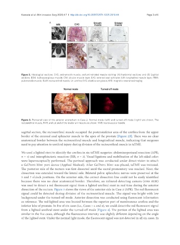
Figure 2. Histological sections. EVG, anti-smooth muscle, and anti-striated muscle staining: (A) horizontal sections; and (B) Sagittal
sections. BSM: bulbospongiosus muscle; CM: circular muscle layer; EAS: external anal sphincter; LM: longitudinal muscle layer; PRM:
puborectalis muscle; RUM: rectourethral muscle; Ur: urethra; EVG: elastica van gieson; MRI: magnetic resonance imaging
Figure 3. Transanal view of the anterior anorectum in Case 2. Normal mode (left) and turned-off mode (right) are shown. The
rectourethral muscle, NVB, and cut end of the levator ani muscle are shown. NVB: neurovascular bundle
sagittal section, the rectourethral muscle occupied the posteroinferior area of the urethra from the upper
border of the external anal sphincter muscle to the apex of the prostate [Figure 2B]. There was no clear
anatomical border between the rectourethral muscle and longitudinal muscle, indicating that surgeons
need to pay attention to urethral injury during division of the rectourethral muscle in taTME.
We used a lighted stent to identify the urethra in six taTME surgeries: abdominoperineal resection (APR;
n = 4) and intersphincteric resection (ISR; n = 2). Vessel ligations and mobilization of the left-sided colon
were laparoscopically performed. The perineal approach was conducted under direct vision to attach
a GelPoint Mini port device (Applied Medical). After GelPoint Mini was placed, taTME was initiated.
The posterior side of the rectum was first dissected until the sacral promontory was reached. Next, the
dissection was extended toward the lateral side. Bilateral pelvic splanchnic nerves were preserved at the
5 and 7 o’clock positions. On the anterior side, the correct dissection line could not be easily identified
because there was no clear anatomical border. Therefore, an infrared-detecting camera (1588 AIM)
was used to detect a red fluorescent signal from a lighted urethral stent in real time during the anterior
dissection of the rectum. Figure 3 shows the views of the anterior side in Case 2 (APR). The red fluorescent
signal could be detected during division of the rectourethral muscle. The signal was bright with low
background under the turned-off mode. Anterior dissection was conducted using fluorescent information
as reference. The red-lighted area was located between the superior part of membranous urethra and the
inferior lobe of prostate. In five of six cases (i.e., Cases 1-4 and 6), we could detect the red fluorescent signal
from a lighted urethral stent under the turned-off mode [Figure 4]. The portion of the lighted area was
similar in the five cases, although the fluorescence intensity was slightly different depending on the angle
of the lighted stent. Under the normal light mode, the fluorescent signal was not detected in all six cases. In