
Page 51 - Read Online
P. 51
Page 2 of 6 Kawada et al. Mini-invasive Surg 2020;4:7 I http://dx.doi.org/10.20517/2574-1225.2019.44
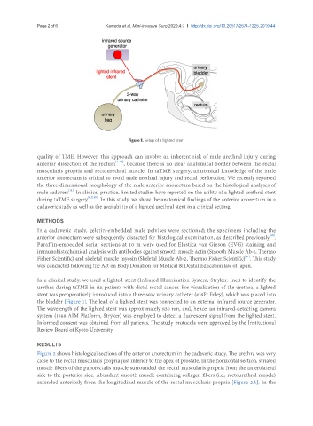
Figure 1. Setup of a lighted stent
quality of TME. However, this approach can involve an inherent risk of male urethral injury during
anterior dissection of the rectum [6-10] , because there is no clear anatomical border between the rectal
muscularis propria and rectourethral muscle. In taTME surgery, anatomical knowledge of the male
anterior anorectum is critical to avoid male urethral injury and rectal perforation. We recently reported
the three-dimensional morphology of the male anterior anorectum based on the histological analyses of
[11]
male cadavers . In clinical practice, limited studies have reported on the utility of a lighted urethral stent
during taTME surgery [6,7,10] . In this study, we show the anatomical findings of the anterior anorectum in a
cadaveric study as well as the availability of a lighted urethral stent in a clinical setting.
METHODS
In a cadaveric study, gelatin-embedded male pelvises were sectioned; the specimens including the
anterior anorectum were subsequently dissected for histological examination, as described previously .
[11]
Paraffin-embedded serial sections at 10 m were used for Elastica van Gieson (EVG) staining and
immunohistochemical analysis with antibodies against smooth muscle actin (Smooth Muscle Ab-1, Thermo
[11]
Fisher Scientific) and skeletal muscle myosin (Skeletal Muscle Ab-2, Thermo Fisher Scientific) . This study
was conducted following the Act on Body Donation for Medical & Dental Education law of Japan.
In a clinical study, we used a lighted stent (Infrared Illumination System, Stryker. Inc.) to identify the
urethra during taTME in six patients with distal rectal cancer. For visualization of the urethra, a lighted
stent was preoperatively introduced into a three-way urinary catheter (#18Fr Foley), which was placed into
the bladder [Figure 1]. The lead of a lighted stent was connected to an external infrared source generator.
The wavelength of the lighted stent was approximately 830 nm, and, hence, an infrared-detecting camera
system (1588 AIM Platform, Stryker) was employed to detect a fluorescent signal from the lighted stent.
Informed consent was obtained from all patients. The study protocols were approved by the Institutional
Review Board of Kyoto University.
RESULTS
Figure 2 shows histological sections of the anterior anorectum in the cadaveric study. The urethra was very
close to the rectal muscularis propria just inferior to the apex of prostate. In the horizontal section, striated
muscle fibers of the puborectalis muscle surrounded the rectal muscularis propria from the anterolateral
side to the posterior side. Abundant smooth muscle containing collagen fibers (i.e., rectourethral muscle)
extended anteriorly from the longitudinal muscle of the rectal muscularis propria [Figure 2A]. In the