
Page 450 - Read Online
P. 450
Page 4 of 9 Scotti et al. Mini-invasive Surg 2020;4:49 I http://dx.doi.org/10.20517/2574-1225.2020.38
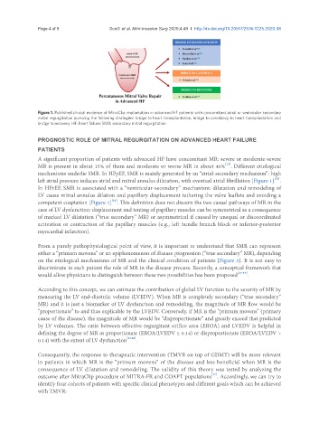
Figure 1. Published clinical evidence of MitraClip implantation in advanced HF patients with concomitant atrial or ventricular secondary
mitral regurgitation pursuing the following strategies: bridge to heart transplantation, bridge to candidacy to heart transplantation and
bridge to recovery. HF: heart failure; SMR: secondary mitral regurgitation
PROGNOSTIC ROLE OF MITRAL REGURGITATION ON ADVANCED HEART FAILURE
PATIENTS
A significant proportion of patients with advanced HF have concomitant MR: severe or moderate-severe
[15]
MR is present in about 15% of them and moderate or worse MR in about 40% . Different etiological
mechanisms underlie SMR. In HFpEF, SMR is mainly generated by an “atrial-secondary mechanism”: high
[16]
left atrial pressure induces atrial and mitral annulus dilatation, with eventual atrial fibrillation [Figure 1] .
In HFrEF, SMR is associated with a “ventricular-secondary” mechanism: dilatation and remodeling of
LV cause mitral annulus dilation and papillary displacement tethering the valve leaflets and avoiding a
[16]
competent coaptation [Figure 1] . This definition does not discern the two casual pathways of MR in the
case of LV dysfunction: displacement and tenting of papillary muscles can be symmetrical as a consequence
of marked LV dilatation (“true secondary” MR) or asymmetrical if caused by unequal or discoordinated
activation or contraction of the papillary muscles (e.g., left bundle branch block or inferior-posterior
myocardial infarction).
From a purely pathophysiological point of view, it is important to understand that SMR can represent
either a “primum movens” or an epiphenomenon of disease progression (“true secondary” MR), depending
on the etiological mechanisms of MR and the clinical condition of patients [Figure 2]. It is not easy to
discriminate in each patient the role of MR in the disease process. Recently, a conceptual framework that
would allow physicians to distinguish between these two possibilities has been proposed [17-19] .
According to this concept, we can estimate the contribution of global LV function to the severity of MR by
measuring the LV end-diastolic volume (LVEDV). When MR is completely secondary (“true secondary”
MR) and it is just a biomarker of LV dysfunction and remodeling, the magnitude of MR flow would be
“proportionate” to and thus explicable by the LVEDV. Conversely, if MR is the “primum movens” (primary
cause of the disease), the magnitude of MR would be “disproportionate” and greatly exceed that predicted
by LV volumes. The ratio between effective regurgitant orifice area (EROA) and LVEDV is helpful in
defining the degree of MR as proportionate (EROA/LVEDV ≤ 0.14) or disproportionate (EROA/LVEDV >
0.14) with the extent of LV dysfunction [17,20] .
Consequently, the response to therapeutic intervention (TMVR on top of GDMT) will be more relevant
in patients in which MR is the “primum movens” of the disease and less beneficial when MR is the
consequence of LV dilatation and remodeling. The validity of this theory was tested by analyzing the
[17]
outcome after MitraClip procedure of MITRA-FR and COAPT populations . Accordingly, we can try to
identify four cohorts of patients with specific clinical phenotypes and different goals which can be achieved
with TMVR: