
Page 37 - Read Online
P. 37
Ohmura et al. Mini-invasive Surg 2019;3:4 I http://dx.doi.org/10.20517/2574-1225.2018.69 Page 3 of 13
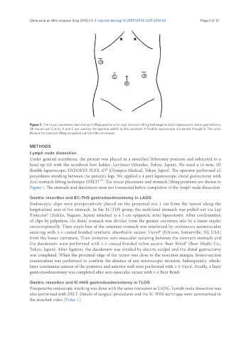
Figure 1. The trocar placement and stomach lifting positions for dual stomach-lifting technique in total laparoscopic distal gastrectomy.
All trocars are 12 mm. A and C are used by the operator and D by the assistant. A flexible laparoscope is inserted through B. The nylon
threads for stomach lifting are pulled out from the star marks
METHODS
Lymph node dissection
Under general anesthesia, the patient was placed in a modified lithotomy position and subjected to a
head-up tilt with the semiboot foot holder: Levitator (Mizuho, Tokyo, Japan). We used a 10-mm, 3D
flexible laparoscope; ENDOEYE FLEX 3D® (Olympus Medical, Tokyo, Japan). The operator performed all
procedures standing between the patient’s legs. We applied a 4 port-laparoscopic distal gastrectomy with
[15]
dual stomach-lifting technique (DSLT) . The trocar placement and stomach lifting positions are shown in
Figure 1. The stomach and duodenum were not transected before completion of the lymph node dissection.
Gastric resection and EC-THS gastroduodenostomy in LADG
Endoscopic clips were preoperatively placed on the proximal site 1 cm from the tumor along the
longitudinal axis of the stomach. In the EC-THS group, the mobilized stomach was pulled out via Lap
Protector® (Hakko, Nagano, Japan) attached to a 5-cm epigastric mini-laparotomy. After confirmation
of clips by palpation, the distal stomach was divided from the greater curvature side by a linear stapler
extracorporeally. Then staple line of the remnant stomach was reinforced by continuous seromuscular
suturing with 3-0 coated braided synthetic absorbable suture: Vicryl® (Ethicon, Somerville, NJ, USA)
from the lesser curvature. Then posterior sero-muscular suturing between the remnant stomach and
the duodenum were performed with 3-0 coated braided nylon suture: Bear Braid® (Bear Medic Co.,
Tokyo, Japan). After ligation, the duodenum was divided by electric scalpel and the distal gastrectomy
was completed. When the proximal edge of the tumor was close to the resection margin, frozen-section
examination was performed to confirm the absence of any microscopic invasion. Subsequently, whole-
layer continuous sutures of the posterior and anterior wall were performed with 3-0 Vicryl. Finally, 2-layer
gastroduodenostomy was completed after sero-muscular suture with 3-0 Bear Braid.
Gastric resection and IC-HHS gastroduodenostomy in TLDG
Preoperative endoscopic marking was done with the same maneuver as LADG. Lymph node dissection was
also performed with DSLT. Details of surgical procedures and the IC-HHS technique were summarized in
the attached video [Video 1].