
Page 282 - Read Online
P. 282
Page 6 of 11 Serra-Aracil et al. Mini-invasive Surg 2019;3:37 I http://dx.doi.org/10.20517/2574-1225.2019.36
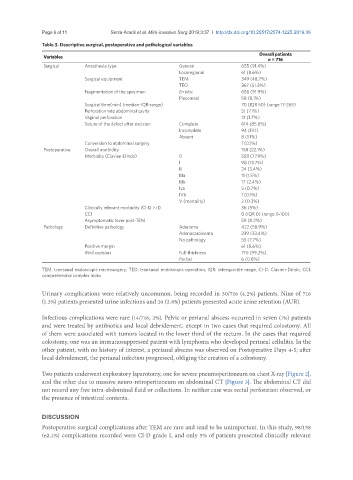
Table 3. Descriptive surgical, postoperative and pathological variables
Overall patients
Variables
n = 716
Surgical Anesthesia type General 655 (91.4%)
Locoregional 61 (8.6%)
Surgical equipment TEM 349 (48.7%)
TEO 367 (51.3%)
Fragmentation of the specimen En bloc 658 (91.9%)
Piecemeal 58 (8.1%)
Surgical time(min) (median-IQR-range) 70 (IQR 50) (range 17-265)
Perforation into abdominal cavity 51 (7.1%)
Vaginal perforation 12 (1.7%)
Suture of the defect after excision Complete 614 (85.8%)
Incomplete 94 (13.1)
Absent 8 (1.1%)
Conversion to abdominal surgery 1 (0.1%)
Postoperative Overall morbidity 158 (22.1%)
Morbidity (Clavien-Dindo) 0 558 (77.9%)
I 98 (13.7%)
II 24 (3.4%)
IIIa 11 (1.5%)
IIIb 17 (2.4%)
Iva 5 (0.7%)
IVb 1 (0.1%)
V (mortality) 2 (0.3%)
Clinically relevant morbidity (Cl-D > II) 36 (5%)
CCI 0 (IQR 0) (range 0-100)
Asymptomatic fever post-TEM 59 (8.2%)
Pathology Definitive pathology Adenoma 422 (58.9%)
Adenocarcinoma 239 (33.4%)
No pathology 55 (7.7%)
Positive margin 61 (8.6%)
Wall excision Full-thickness 710 (99.2%)
Partial 6 (0.8%)
TEM: transanal endoscopic microsurgery; TEO: transanal endoscopic operation; IQR: interquartile range; Cl-D: Clavien-Dindo; CCI:
comprehensive complex index
Urinary complications were relatively uncommon, being recorded in 30/716 (4.2%) patients. Nine of 716
(1.3%) patients presented urine infections and 20 (2.8%) patients presented acute urine retention (AUR).
Infectious complications were rare (14/716, 2%). Pelvic or perianal abscess occurred in seven (1%) patients
and were treated by antibiotics and local debridement, except in two cases that required colostomy. All
of them were associated with tumors located in the lower third of the rectum. In the cases that required
colostomy, one was an immunosuppressed patient with lymphoma who developed perineal cellulitis. In the
other patient, with no history of interest, a perianal abscess was observed on Postoperative Days 4-5; after
local debridement, the perianal infection progressed, obliging the creation of a colostomy.
Two patients underwent exploratory laparotomy, one for severe pneumoperitoneum on chest X-ray [Figure 2],
and the other due to massive neuro-retroperitoneum on abdominal CT [Figure 3]. The abdominal CT did
not record any free intra-abdominal fluid or collections. In neither case was rectal perforation observed, or
the presence of intestinal contents.
DISCUSSION
Postoperative surgical complications after TEM are rare and tend to be unimportant. In this study, 98/158
(62.1%) complications recorded were Cl-D grade I, and only 5% of patients presented clinically relevant