
Page 145 - Read Online
P. 145
de Pascale et al. Mini-invasive Surg 2019;3:18 I http://dx.doi.org/10.20517/2574-1225.2019.04 Page 3 of 10
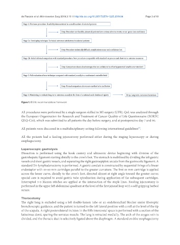
Figure 1. IDEAL recommendations framework
All procedures were performed by a single surgeon skilled in MI surgery (UFR). QoL was analyzed through
the European Organization for Research and Treatment of Cancer Quality of Life Questionnaire (EORTC
QLQ-C30), which was submitted to all patients the day before surgery, and at postoperative day 7 and 90.
All patients were discussed in a multidisciplinary setting following international guidelines .
[7]
All the patients had a feeding jejunostomy performed either during the staging laparoscopy or during
esophagectomy.
Laparoscopic gastrolysis
Dissection is performed using the hook cautery and ultrasonic device beginning with division of the
gastrohepatic ligament starting distally to the crow’s foot. The stomach is mobilized by dividing the left gastric
vessels and short gastric vessels, and separating the right gastroepiploic arcade from the gastrocolic ligament. A
standard D2-lymphadenectomy is performed. A gastric conduit is constructed by sequential firings of a linear
endostapler with 45-60 mm cartridges parallel to the greater curvature. The first 45 mm cartridge is applied
across the lesser curve, distally to the crow’s foot, directed almost at right angle toward the greater curve;
special care is required to avoid gastric tube spiralization during application of the subsequent cartridges.
Interrupted 3-0 Maxon stitches are applied at the intersection of the staple lines. Feeding jejunostomy is
performed in the upper left abdominal quadrant at the level of the first jejunal loop with a self-gripping barbed
suture.
Thoracotomy
The right lung is excluded using a left double-lumen tube or an endobronchial blocker under fiberoptic
bronchoscopic guidance, and the patient is turned to the left lateral position with a roll at the level of the tip
of the scapula. A right posterolateral incision in the fifth intercostal space is performed with a section of the
latissimus dorsi, sparing the serratus muscle. The lung is retracted medially. The arch of the azygos vein is
divided, and the thoracic duct is selectively ligated above the diaphragm. A standard en-bloc esophagectomy