
Page 140 - Read Online
P. 140
Goh et al. Mini-invasive Surg 2019;3:17 I http://dx.doi.org/10.20517/2574-1225.2019.06 Page 3 of 5
A
B
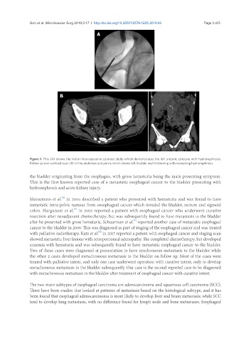
Figure 1. This (A) shows the initial intra-operative contrast study which demonstrates the left ureteric stricture with hydronephrosis.
Follow up non contrast scan (B) of the abdomen and pelvis which shows left bladder wall thickening with worsening hydronephrosis
the bladder originating from the esophagus, with gross hematuria being the main presenting symptom.
This is the first known reported case of a metastatic esophageal cancer to the bladder presenting with
hydronephrosis and acute kidney injury.
[5]
Matsumoto et al. in 2004 described a patient who presented with hematuria and was found to have
metastatic intra-pelvic tumour from oesophageal cancer which invaded the bladder, rectum and sigmoid
colon. Hargunani et al. in 2005 reported a patient with esophageal cancer who underwent curative
[6]
resection after neoadjuvant chemotherapy; but was subsequently found to have metastasis to the bladder
after he presented with gross hematuria. Schuurman et al. reported another case of metastatic esophageal
[7]
cancer to the bladder in 2009. This was diagnosed as part of staging of the esophageal cancer and was treated
[8]
with palliative radiotherapy. Katz et al. in 2017 reported a patient with esophageal cancer and staging scan
showed metastatic liver lesions with retroperitoneal adenopathy. She completed chemotherapy, but developed
anaemia with hematuria and was subsequently found to have metastatic esophageal cancer to the bladder.
Two of these cases were diagnosed at presentation to have synchronous metastasis to the bladder while
the other 2 cases developed metachronous metastasis to the bladder on follow up. Most of the cases were
treated with palliative intent, and only one case underwent operation with curative intent, only to develop
metachronous metastasis to the bladder subsequently. Our case is the second reported case to be diagnosed
with metachronous metastasis to the bladder after treatment of esophageal cancer with curative intent.
The two main subtypes of esophageal carcinoma are adenocarcinoma and squamous cell carcinoma (SCC).
There have been studies that looked at patterns of metastasis based on the histological subtype, and it has
been found that esophageal adenocarcinoma is more likely to develop liver and brain metastasis; while SCC
tend to develop lung metastasis, with no difference found for lymph node and bone metastases. Esophageal