
Page 158 - Read Online
P. 158
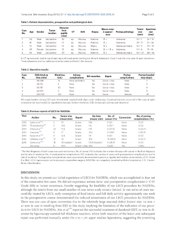
Toma et al. Mini-invasive Surg 2018;2:21  I  http://dx.doi.org/10.20517/2574-1225.2018.24                                           Page 5 of 8

               Table 1. Patient characteristics, preoperative and pathological data
                                         Distance                     Macro-scop-             Tumor  Specimen
                Case  Age   Gender   Tumor   from   CT   EUS   Biopsy   ic appear-  Postop pathology   size  size
                No.               location   papilla                    ance                  (mm)   (mm)
                                          (mm)
                1    54   Male   2nd portion     32   np   Mucosa   Adenma   Ⅱ c   Adenoma   12 × 7   22 × 15
                2    41   Male   2nd portion      15   np   Mucosa   Adenma   Ⅱ c   Adenoma   21 × 17   31 × 26
                3    57   Male   2nd portion      14   np   Mucosa   Atypia   Ⅱ a   Adeno-carcinoma   16 × 9   29 × 19
                4    69   Female  2nd portion      21   np   Mucosa   Adenma   Ⅱ c + Ⅱ a   Adenoma   14 × 6   19 × 14
                5    50   Male   2nd portion     29   np   Mucosa   Adenma   Ⅱ a   Adeno-carcinoma   54 × 47   68 × 55
               In CT, np indicates neither significant regional lymph node swelling nor distant metastasis. Case 5 was the only case of open conversion.
               Three adenomas and two adenocarcinomas were confined in the mucosa


               Table 2. Operative results
                Case     ESD/total op   Blood loss   Intraop                         Postop    Postop hospital
                No.      time (min)     (mL)    complications   R0-resection   Repair   complications   stay (days)
                1         98/228        10      Micro-perforation   Yes   Suture + clips   None     7
                2         49/152        5       None           Yes    Suture + clips   None         9
                3         56/187        30      None           Yes    Suture + clips   None         8
                4         26/202        5       None           Yes    Suture + clips   None         8
                5         252/552       100     None           Yes    Suture         Stenosis       49
               Micro-perforation during ESD was immediately repaired with clips under endoscopy. Duodenal stenosis occurred in the case of open
               conversion but was treated by repeated endoscopic balloon dilatation. ESD: endoscopic submucosal dissection


               Table 3. Previous reports of LECS for NADENs
                Year                      Tumor size           Op time    No. of             No. of postop
                        Author       No.              Repair                      Conversion
                                         (mean:mm)            (mean; min)  cancer (%)       complications (%)
                2010   Sakon et al. [10]   2   19   Suture   168          0 (0)    None        0 (0)
                               [11]
                2010   Tsujimoto et al.    2   21.5   Suture   116        0 (0) *   None       0 (0)
                             [12]
                2014   Ohata et al.    22   13.3    Suture   133          6 (27.3)   None      5 (22.7)
                            [13]
                2015   Irino et al.    3   17       Suture   234          3 (100)   None       1 (33.3)
                2015   Kyuno et al. [14]    2   12.5   Suture   131       0 (0)    None        0 (0)
                              [15]
                2016   Ichikawa et al.    12   22   Suture   322          10 (83.3)   None     2 (16.7)
                2018   Ojima et al. [20]    18   18 (median)   Suture   116.5 (median)   4 (38.9)   None   0 (0)
                     Our study       5    23.4      Suture + clips  264   2 (40)   Yes         1 (20)
               *The final diagnosis of both cases was carcinoid tumors. No. of cancer (%) indicates the number of cases with cancer in the final diagnosis
               and its rate of incidence. No. of postoperative complications (%) indicates the number of cases with postoperative complications and its
               rate of incidence. Postoperative complications were occasionally documented in previous reports, but treated conservatively (C-D: Grade
               II or IIIa). LECS: laparoscopic and endoscopic cooperative surgery; NADENs: non-ampullary duodenal epithelial neoplasms; C-D: Clavien-
               Dindo Classification


               DISCUSSION
               In this study, we present our initial experience of LECS for NADENs, which was accomplished in four out
               of the consecutive five cases. We did not experience serious intra- and postoperative complications (> C-D:
               Grade IIIb) or tumor recurrence, thereby suggesting the feasibility of our LECS procedure for NADENs,
               although the results from our small number of case series study remain limited. In our series of cases suc-
               cessfully treated by LECS, early resumption of food intake and full daily activity approximately one week
               in the postoperative course demonstrated the reduced invasiveness of our LECS procedure for NADENs.
               There was one case of open conversion due to the relatively large mucosal defect (tumor size: 54 mm ×
               47 mm in case 5) resulting from ESD in this study, implying the limitation of the indication of our proce-
                                                [17]
               dure for LECS for NADENs. Abe et al.  reported the successful treatment of duodenal GIST, 62 mm in di-
               ameter by laparoscopy-assisted full-thickness resection, where both resection of the lesion and subsequent
               repair was performed manually under the 5 to 7 cm upper median laparotomy, suggesting the promising