
Page 156 - Read Online
P. 156
Toma et al. Mini-invasive Surg 2018;2:21 I http://dx.doi.org/10.20517/2574-1225.2018.24 Page 3 of 8
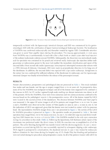
Figure 1. Port site. The value in the circle indicates the size of each port inserted in the abdomen
temporarily occluded with the laparoscopic intestinal clamper, and ESD was commenced by the gastro-
enterologist (KH with the certification of Japan Gastroenterological Endoscopy Society). The localization
of NADENs was confirmed endoscopically and thereafter treated by regular ESD. Considerable attention
was given to avoid Vater papilla injury during the procedure. The mucosa approximately 2-3 mm away
from NADENs was circumferentially incised with either a flush knife or clutch cutter after local injection
of the sodium hyaluronate solution around NADENs. Subsequently, the dissection was meticulously made,
and the specimen was contained in the pouch and retrieved orally. Endoscopic dye injection (either indi-
gocarmine or indocyanine green) to the ulcer bed enabled the immediate identification and repair of the
mucosal defect from serosal side under laparoscopy. Intracorporeal-interrupted seromuscular sutures with
4-0 PDS (Johnson & Johnson, Cincinnati, OH) were made in perpendicular direction to the long axis of
the duodenum. In addition, the mucosal defect was repaired with clips under endoscopy. Airtightness in
the suture line was confirmed by sufficient inflation of the duodenum by endoscopy, and the laparoscopic
intestinal clamper was finally retrieved before the release of the pneumoperitoneum.
RESULTS
Patient characteristics, preoperative and pathological data are presented in Table 1. This study included
four males and one female, and the age at surgery ranged from 41 to 69 years old. In preoperative data,
none of the five NADENs was malignant in biopsy and all of the lesions were supposed to be confined in
the mucosa in EUS. In CT, neither regional lymph node swelling nor distant metastasis was shown in all
of the patients. Of the five NADENs, there were three adenomas and two adenocarcinomas confined in the
mucosa based on the pathological reports in the postoperative course. All of the lesions treated by LECS
were located in the second portion of the duodenum. The distance between NADENs and Vater papilla
was measured in the upper-GI series images in all of the patients and ranged from 14 to 32 mm. In cases
2 and 3, NADENs were observed in the vicinity of Vater papilla (15 mm in case 2, 14 mm in case 3), but
the indication of LECS was approved, given that the lesions were not located in contact with the pancreas
head. The representative NADENs images from case 3 are presented in Figure 2. The section of the LECS
for NADENs procedure is presented in Figure 3. The operative results are presented in Table 2. The total
operation time ranged from 152 to 552 (mean 264) min. In case 5, the relatively large mucosal defect result-
ing from ESD (tumor size: 54 mm × 47 mm in Table 1) for NADENs resulted in the only open conversion
during the procedure in this study [Table 2]. Following the laparotomy, full thickness suture repair of the
duodenum was performed under the direct vision. In ESD, R0-resection was accomplished in all of the
patients. The active bleeding during ESD was mainly treated with the electrocoagulation. In case 1, micro-
perforation occurred during ESD, but was successfully repaired by subsequent clipping under endoscopy.
With regard to postoperative complications, the duodenal stenosis in case 5 was treated by endoscopic
balloon dilatation for four times during the hospital stay (C-D: Grade IIIa), but bleeding and perforation
were not noted during the procedure and postoperative course. In all of the patients treated with LECS for