
Page 141 - Read Online
P. 141
Basso et al. Mini-invasive distal pancreatectomy
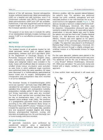
tumours of the left pancreas. Several retrospective lithotomic position, with the operator placed between
studies confirmed laparoscopic distal pancreatectomy the patient’s legs. The operation was performed
(LDP) as a feasible and safe technique, even if no through four ports: umbilical, subxyphoid, and both
randomized controlled trials (RCTs) comparing open subcostal positions in the mid-clavicular line so as to
distal pancreatectomy (ODP) and LDP are available. avoid trauma to the epigastric vessels [Figure 1]. The
Furthermore, it has been argued that costs for reduced devices included a harmonic scalpel (Harmonic ACE ,
®
hospital length of stay (LoS) are counterbalanced by Ethicon EndoSurgery, Cincinnati, OH, USA) used
the increased operative costs of LDP. [7] for dissection. Intraoperative ultrasound was used if
needed to localize the tumour. In cases without splenic
The purpose of our study was to evaluate the safety preservation, a vascular stapler was used to divide
of our standardized minimally invasive technique and the splenic vein and two Hem-o-lok (Teleflex Medical
®
assess if LDP is a cost-effective procedure compared Europe Ltd., IDA Business and Technology Park,
to ODP. Athlone, Ireland) clips were applied on the splenic
artery. Division of the pancreas was performed using
METHODS a stapler. The specimen was placed in an Endopouch
Retrieval Bag (Ethicon EndoSurgery, Cincinnati, OH,
®
Study design and population USA) and removed through a slightly enlarged peri-
The medical records of all patients treated for left- umbilical incision or a Pfannenstiel incision for large
sided pancreatic lesions (with or without splenic specimens.
preservation), between April 2013 and March 2015,
at the Department of Oncologic Surgery at the For the open approach, patients were placed in the
Humanitas Gavazzeni Institute of Bergamo (Italy), supine position and a left subcostal incision was used.
were retrospectively analysed. Patients with both The additional cost for the use of Harmonic Focus
benign and malignant lesions were included in the + Long Shears (Ethicon EndoSurgery, Cincinnati,
®
study. Cases with insufficient data for analysis or that OH, USA) was calculated. The pancreatic stump was
entailed simple tumour enucleation were excluded, treated with a stapler, similarly to the laparoscopic
as were those in which additional organ resections approach.
were performed during the same operation. All cases
were discussed in a multidisciplinary gastrointestinal A close suction drain was placed in all cases and
tumour board prior to surgery. Demographics and
intraoperative and postoperative data were recorded
in an ad hoc database.
The American Society of Anaesthesiologists (ASA)
score was reported, and body mass index (BMI) was
[8]
calculated for each patient. Intraoperative blood loss,
operative time, hospital LoS, postoperative morbidity,
perioperative mortality (within 30 days from surgery),
and 30-day readmission rates were also recorded.
The level of pain reported was recorded three times
per day on postoperative days 1 and 2, using the
standard visual analogic scale (VAS). The presence
of a postoperative pancreatic fistula was assessed
according to the 2005 International Study Group on
Pancreatic Fistula (ISGPF) criteria. Analysis of costs
[9]
included the expenses for the hospital stay, operative
time and equipment (surgical staplers and energy
devices), pharmaceutical treatment, nursing, and
laboratory and pathology fees. No post-discharge care
or home-nursing costs were included.
Surgical technique
All pancreatic resections were performed by Figure 1: Position of trocar sites. (1) 10/12 mm umbilicus; (2)
10/12 mm left anterior axillary line between the costal margin and
experienced surgeons using a standardized the iliac crest; (3) 5 mm subxiphoid area; (4) 5 mm lateral right
technique. The LDP patients were placed in the rectus sheath under the right costal margin
134 Mini-invasive Surgery ¦ Volume 1 ¦ September 30