
Page 11 - Read Online
P. 11
Koga Improved PETA for LFS at L5/S1
A C E G I
B D F H J
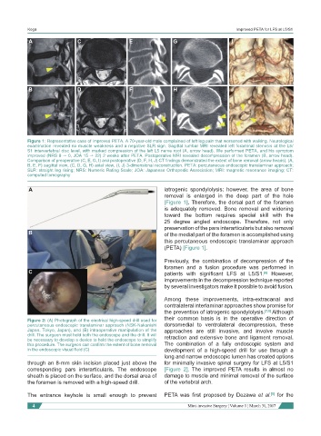
Figure 1: Representative case of improved PETA. A 70-year-old male complained of left leg pain that worsened with walking. Neurological
examination revealed no muscle weakness and a negative SLR sign. Sagittal lumbar MRI revealed left foraminal stenosis at the L5/
S1 intervertebral disc level, with marked compression of the left L5 nerve root (A, arrow head). We performed PETA, and his symptom
improved (NRS 8 → 0, JOA 15 → 22) 2 weeks after PETA. Postoperative MRI revealed decompression of the foramen (B, arrow head).
Comparison of preoperative (C, E, G, I) and postoperative (D, F, H, J) CT findings demonstrated the extent of bone removal (arrow heads). (A,
B, E, F) sagittal view, (C, D, G, H) axial view, (I, J) 3-dimensional reconstruction. PETA: percutaneous endoscopic translaminar approach;
SLR: straight leg rising; NRS: Numeric Rating Scale; JOA: Japanese Orthopedic Association; MRI: magnetic resonance imaging; CT:
computed tomography
A iatrogenic spondylolysis; however, the area of bone
removal is enlarged in the deep part of the hole
[Figure 1]. Therefore, the dorsal part of the foramen
is adequately removed. Bone removal and widening
toward the bottom requires special skill with the
25 degree angled endoscope. Therefore, not only
preservation of the pars interarticularis but also removal
B of the medial part of the foramen is accomplished using
this percutaneous endoscopic translaminar approach
(PETA) [Figure 1].
Previously, the combination of decompression of the
foramen and a fusion procedure was performed in
C patients with significant LFS at L5/S1. However,
[6]
improvements in the decompression technique reported
by several investigators make it possible to avoid fusion.
Among these improvements, intra-extracanal and
contralateral interlaminar approaches show promise for
the prevention of iatrogenic spondylolysis. [7,8] Although
their common basis is in the operative direction of
Figure 2: (A) Photograph of the electrical high-speed drill used for
percutaneous endoscopic translaminar approach (NSK-Nakanishi dorsomedial to ventrolateral decompression, these
Japan, Tokyo, Japan), and (B) intraoperative manipulation of the approaches are still invasive, and involve muscle
drill. The surgeon must hold both the endoscope and the drill. It will retraction and extensive bone and ligament removal.
be necessary to develop a device to hold the endoscope to simplify
this procedure. The surgeon can confirm the extent of bone removal The combination of a fully endoscopic system and
in the endoscopic visual field (C) development of a high-speed drill for use through a
long and narrow endoscopic lumen has created options
through an 8-mm skin incision placed just above the for minimally invasive spinal surgery for LFS at L5/S1
corresponding pars interarticularis. The endoscope [Figure 2]. The improved PETA results in almost no
sheath is placed on the surface, and the dorsal area of damage to muscle and minimal removal of the surface
the foramen is removed with a high-speed drill. of the vertebral arch.
The entrance keyhole is small enough to prevent PETA was first proposed by Dezawa et al. for the
[5]
4 Mini-invasive Surgery ¦ Volume 1 ¦ March 31, 2017