
Page 90 - Read Online
P. 90
Nakamura et al. Mini-invasive Surg 2022;6:50 https://dx.doi.org/10.20517/2574-1225.2022.38 Page 11 of 20
Figure 6. (A) Schema of “trimming”. The trimming is performed until the surface of the muscularis propria. (B) Endoscopic image of
trimming during endoscopic submucosal dissection (ESD). The muscularis propria behind the transparent submucosa is whitish. (C)
After trimming and injection of solution into the submucosa, the initial submucosal plane for submucosal dissection is recognized easily.
Isolation of the specimen from the normal mucosa avoids leakage of the injected solution and allows efficient mucosal lifting by
submucosal injection. (D) After “trimming”, submucosal dissection at the lateral or distal part of the specimen can be easily performed.
Note that the deep submucosa is less vascular and fibrous.
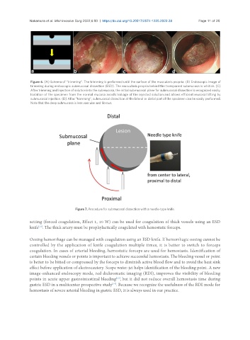
Figure 7. Procedure for submucosal dissection with a needle-type knife.
setting (forced coagulation, Effect 1, 10 W) can be used for coagulation of thick vessels using an ESD
[12]
knife . The thick artery must be prophylactically coagulated with hemostatic forceps.
Oozing hemorrhage can be managed with coagulation using an ESD knife. If hemorrhagic oozing cannot be
controlled by the application of knife coagulation multiple times, it is better to switch to forceps
coagulation. In cases of arterial bleeding, hemostatic forceps are used for hemostasis. Identification of
certain bleeding vessels or points is important to achieve successful hemostasis. The bleeding vessel or point
is better to be bitted or compressed by the forceps to diminish active blood flow and to avoid the heat sink
effect before application of electrocautery. Scope water-jet helps identification of the bleeding point. A new
image-enhanced endoscopy mode, red dichromatic imaging (RDI), improves the visibility of bleeding
points in acute upper gastrointestinal bleeding , but it did not reduce overall hemostasis time during
[13]
[14]
gastric ESD in a multicenter prospective study . Because we recognize the usefulness of the RDI mode for
hemostasis of severe arterial bleeding in gastric ESD, it is always used in our practice.