
Page 94 - Read Online
P. 94
Nakamura et al. Mini-invasive Surg 2022;6:50 https://dx.doi.org/10.20517/2574-1225.2022.38 Page 15 of 20
Table 5. Risk factors for Intraoperative perforation of gastric ESD
Risk factors
Location of the lesion (the corpus, the greater curvature, and remnant stomach)
Large (> 20 mm) tumor size
Submucosal tumor invasion or beyond
Submucosal fibrosis
Elevated morphology
Long procedure time
Piecemeal resection
Prior workload of operators
ESD: Endoscopic submucosal dissection.
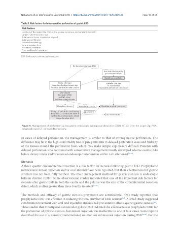
Figure 9. Management of perforation during gastric endoscopic submucosal dissection (ESD). OTSC: Over the scope clip; PGA:
polyglycolic acid; CT: computed tomography.
In cases of delayed perforation, the management is similar to that of intraoperative perforation. The
difference may lie in the high comorbidity rate of pan-peritonitis in delayed perforation cases and friability
of the tissues around the perforation hole, which may make simple clip closure difficult. Patients with
delayed perforation who recovered with conservative management mostly developed adverse events (AE)
before dietary intake and/or received endoscopic intervention within 24 h after onset [73,75] .
Stenosis
A three-quarter circumferential resection is a risk factor for stenosis following gastric ESD. Prophylactic
intralesional steroid injection and/or oral steroids have been reported, but their effectiveness for gastric
stricture has not been fully verified. The main management method for gastric stenosis is endoscopic
balloon dilation (EBD). Some observational studies indicated that one of the important risk factors for
stenosis after gastric ESD in both the cardia and the pylorus was the size of the circumferential resection
defect, which is often greater than three-fourths in extent [81-85] .
The methods and efficacy of gastric stenosis prevention are controversial. One study reported that
[82]
prophylactic EBD was effective in reducing the total number of EBD sessions . A small study suggested
combination treatment with oral and injectable steroids had preventative effects against gastric stenosis .
[86]
Three studies that investigated stenosis after pyloric ESD indicated the effectiveness of prophylactic EBD for
the prevention of pyloric stenosis, but steroid injection was ineffective in one of four cases. Some reports
described the use of a steroid (triamcinolone) solution for submucosal injection during ESD [87,88] . For the