
Page 29 - Read Online
P. 29
De Luca et al. Mini-invasive Surg 2022;6:13 https://dx.doi.org/10.20517/2574-1225.2021.127 Page 5 of 8
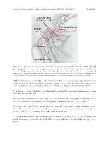
Figure 1. Total anatomical reconstruction technique: the posterior reconstruction is performed in a triple layer by using a 3/0 barbed
suture. The first layer involves Denonvilliers’ fascia and the median raphe, the second the retrotrigonal fascia and the median raphe, and
the third the bladder neck and the rhabdosphincter. The urethra-vesical anastomosis is made by using two 3/0 barbed hemi-running
sutures, involving the full thickness of either the bladder or the urethra. The anterior reconstruction consists of two layers of 3/0 barbed
running sutures. The first layer involves the muscular fibres of the bladder and the peri-urethral tissue. The second involves the vesical
apron and the portion of the endopelvic fascia that covers the dorsal vein complex while involving the pubo-prostatic ligaments.
sRARP was performed successfully in all the cases without the need of conversion. No major intraoperative
complication occurred. All the patients underwent extended lymph node dissection and 3 patients (27.2%)
[21]
could benefit from a monolateral intermediate nerve-sparring according to Pasadena Classification .
No differences in terms of technical surgical difficulties were found between patients undergoing primary
focal or whole-prostate HIFU.
Median catheterization time was 5 days (IQR: 4-7). In 3 patients (27.2%), cystography was performed in the
5th post-operative day, before catheter removal. Median hospital stay was 6 days (IQR: 6-8 days).
Focusing on safety, only Clavien 1 complications were recorded. One patient had postoperative fever treated
with antibiotics infusion, and 1 patient had an asymptomatic lymphocele (about 4 cm in maximum
diameter) detected at an ultrasound performed 3 months post-surgery.
On postoperative histopathology assessment, positive surgical margin rate was 27.2%; in all of these cases,
an extracapsular extension of the disease (≥pT3) was present and none of these patients underwent nerve-
sparring.