
Page 93 - Read Online
P. 93
Page 6 of 11 Avvedimento et al. Mini-invasive Surg 2022;6:24 https://dx.doi.org/10.20517/2574-1225.2021.143
Given these considerations, in redo-TAVI, a tailored screening on the risk of coronary obstruction is
strongly recommended. It should be carefully performed in each patient and achieved through dedicated
pre-procedural CT and fluoroscopic imaging analysis.
ACHIEVING COMMISSURAL ALIGNMENT WITH DIFFERENT THV TYPES
Patient-specific alignment of TAVI devices is nowadays allowed by pre-procedural CT analysis, performed
not only to identify the coplanar implant view but also to add anatomical information about coronary ostia
orientation and native commissures. CT-based commissural alignment can be planned, aiming to identify
specific fluoroscopic projection or predicting TAVI system axial rotation. The Alignment of Transcatheter
Aortic-Valve Neo-Commissures (ALIGN TAVR) study first evaluated the impact of initial THV
deployment orientation on neo-commissural overlap with coronary arteries [19,20] . Afterwards, other studies
have extensively addressed this issue.
In this section, we provide a summary of available data for the main types of TAVI devices.
Self-expanding THVs
The importance of commissural alignment during self-expandable THV implantation is highlighted above.
In the ALIGN TAVR study, 828 TAVR implants (SAPIEN 3, 483; Evolut, 245; ACURATE-neo, 100) were
analyzed using pre-procedural multidetector row CT and coplanar fluoroscopy coregistration. Significantly
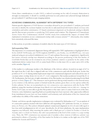
less severe coronary overlap was obtained according to the precise positioning of the Hat marker of
CoreValve Evolut that can be oriented in one of four positions: anterior or posterior in the center of the
deployment device [center front (CF) or center back (CB)] or in the inner (IC) or outer curve (OC) of the
aortic annulus [Figure 4].
A Hat marker is a radiopaque marker at the distal part of the delivery catheter, located at an angle of 90° to
the right from the C-tab, that is aligned with one of the commissures and with the flush port. Evolut Hat
position at OC or CF during initial deployment improved commissural alignment and reduced the rate of
coronary artery overlap (from 36% to 60%; P < 0.05) compared to Hat marker positioned toward the IC or
CB. Furthermore, the best rate of right orientation of the Hat marker was obtained positioning Evolut flush
[21]
port at 3 o’clock [Figure 5] . This maneuver can be performed during initial insertion in the femoral artery
as the rotation of the delivery system is limited inside the patient, given there are two spines within the
Evolut catheter. In fact, we accumulated over 450 consecutive case experiences with the Evolut THV
platform using this insertion technique from March 2019 and from January 2020 to July 2021. Among 316
Evolut TAVI cases, we were able to get the Hat position to the outer curve or center front 96.8% of the time.
However, some limitations regarding flush port positioning have been reported: other approaches different
from transfemoral access have not been studied, and tortuosity and calcification of the ileo-femoral axis
could reduce the ability of Hat marker orientation.
[22]
Patient- and device-specific implantation techniques were evaluated by Bieliauskas et al. in an analysis of
60 consecutive patients treated with three different self-expanding THV platforms (Evolut, 20; Acurate-
neo2, 20; Portico, 20). The authors obtained optimal commissural alignment in 60% of cases with equal or
less than mild misalignment in 88% of cases. Specifically, for all three platforms, the same implantation
sequence was followed. After inserting the system with a flush port at 3 o’clock for Evolut, 6 o’clock for
Acurate, and 12 o’clock for Portico, the valve was positioned at the level of the anulus in a cusp overlap
view. Then, the THV orientation was assessed and, if needed, a slow rotation of delivery catheter was
performed to obtain a CF position of the Hat marker for Evolut or to isolate on the right one of the THV
markers for Acurate and Portico .
[22]