
Page 81 - Read Online
P. 81
Page 10 of 16 Annibali et al. Mini-invasive Surg 2022;6:12 https://dx.doi.org/10.20517/2574-1225.2021.101
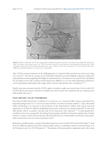
Figure 3. TAVR-in-TAVR after ViV TAVR. A degenerated Carpentier-Edwards Perimount 27 mm bioprosthesis (Edwards Lifesciences,
Irvine, California, USA) treated with a first TAVR CoreValve Evolut R 29 mm (Medtronic, Minneapolis, Minnesota, USA) and a
subsequent Sapien3 Ultra 26 mm (Edwards Lifesciences, Irvine, California, USA), due to damage to CoreValve leaflets after post-
dilation by incomplete expansion with severe aortic regurgitation.
After TAVR, coronary treatment can be challenging, but it is reported with a good success rate in more than
[59]
90% of cases . The risk of coronary artery obstruction should be assessed similarly to that of a surgical BV
with particular caution regarding STJ height as mentioned above. Placement of a second TAVR, especially if
SE, can make access to the coronary arteries much more difficult, so it is even more important to perform a
correct implantation that respects the origin of the coronary ostia and the valve commissures [13,73] .
Finally, long-term outcome from the VIVID registry revealed an eight-year survival rate of 38.0% after ViV
TAVR with the main factors related to mortality and reintervention were small true ID, pre-existing severe
[74]
PPM and BE valve use .
POST-IMPLANT VALVE THROMBOSIS
Subclinical leaflet thrombosis is defined as the presence of a reduced leaflet motion associated with
hypoattenuating lesions on CT and an increased number of transient ischemic attacks . Hypo-attenuated
[75]
leaflet thickening is an increase in the thickness of the bioprosthetic leaflets with typical meniscal
appearance in at least two different multiplanar projections, evidenced on contrast-enhanced CT scan, with
still unclear effects on patient outcome and on the long-term valve function [76,77] . A semi-quantitative
classification has been assumed by describing the percentage of leaflet involvement starting from its basal
insertion. Causes of leaflet thickening and reduced leaflet motion include leaflet thrombosis, endocarditis,
[6]
leaflet deterioration and valve frame expansion issues .
Reduction in leaflets motion caused by thrombosis has been noted in both TAVR and SAVR valves . Data
[78]
from the different registries show that reduced leaflet motion is a relatively common event involving 4% of
SAVR patients and 13% of TAVR patients . Thrombosis of the transcatheter-implanted aortic valve rarely
[75]