
Page 43 - Read Online
P. 43
Page 6 of 13 Cangemi et al. Mini-invasive Surg 2022;6:3 https://dx.doi.org/10.20517/2574-1225.2021.99
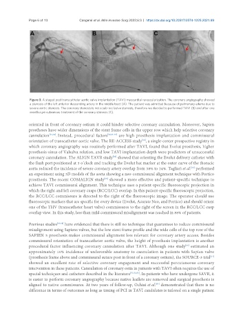
Figure 3. A staged post transcatheter aortic valve implantation (TAVI) myocardial revascularization. The coronary angiography showed
a stenosis of the left anterior descending artery in the middle tract (A). The patient was admitted because of pulmonary edema due to
severe aortic stenosis. The coronary stenosis is not a sub-occlusive stenosis, therefore we decided to performed TAVI (B) and after one
month a percutaneous treatment of the coronary stenosis (C).
oriented in front of coronary ostium it could hinder selective coronary cannulation. Moreover, Sapien
prostheses have wider dimensions of the stent frame cells in the upper row which help selective coronary
cannulation [51,54] . Instead, procedural factors [53,55-57] are high prosthesis implantation and commissural
orientation of transcatheter aortic valve. The RE-ACCESS study , a single-center prospective registry in
[53]
which coronary angiography was routinely performed after TAVI, found that Evolut prosthesis, higher
prosthesis-sinus of Valsalva relation, and low TAVI implantation depth were predictors of unsuccessful
[58]
coronary cannulation. The ALIGN TAVR study showed that orienting the Evolut delivery catheter with
the flush port positioned at 3 o’clock and tracking the Evolut hat marker at the outer curve of the thoracic
aorta reduced the incidence of severe coronary artery overlap from 38% to 24%. Tagliari et al. performed
[59]
an experiment using 3D models of the aorta showing a neo-commissural alignment technique with Portico
prosthesis. The recent COMALIGN study showed a more effective and patient-specific technique to
[60]
achieve TAVI commissural alignment. This technique uses a patient-specific fluoroscopic projection in
which the right and left coronary cusps (RCC/LCC) overlap. In this patient-specific fluoroscopic projection,
the RCC/LCC commissure is directed to the right of the fluoroscopic image. The operator should use
fluoroscopic markers that are specific for every device (Evolut, Acurate Neo, and Portico) and should orient
one of the THV (transcatheter heart valve) commissures to the right of the screen in the RCC/LCC cusp
overlap view. In this study, less than mild commissural misalignment was reached in 88% of patients.
Previous studies [57,58] have evidenced that there is still no technique that guarantees to reduce commissural
misalignment using Sapiens valves, but the low stent frame profile and the wide cells of the top row of the
SAPIEN 3 prosthesis makes commissural alignment less relevant for coronary artery access. Besides
commissural orientation of transcatheter aortic valve, the height of prosthesis implantation is another
procedural factor influencing coronary cannulation after TAVI. Although one study estimated an
[57]
approximately 10% incidence of unfavorable anatomy to cannulation in patients with Sapien valve
(prosthesis frame above and commissural suture post in front of a coronary ostium), the SOURCE-3 trial
[61]
showed an excellent rate of selective coronary engagement and successful percutaneous coronary
intervention in these patients. Cannulation of coronary ostia in patients with TAVI often requires the use of
special techniques and catheters described in the literature [51,62,63] . In patients who have undergone SAVR, it
is easier to perform coronary angiography because native leaflets are removed and surgical prosthesis is
aligned to native commissures. At two years of follow-up, Ochiai et al. demonstrated that there is no
[64]
difference in terms of outcomes as long as timing of PCI in TAVI candidates is tailored on a single patient