
Page 42 - Read Online
P. 42
Cangemi et al. Mini-invasive Surg 2022;6:3 https://dx.doi.org/10.20517/2574-1225.2021.99 Page 5 of 13
Table 1. Timing of PCI in relation to TAVI
Advantages Disadvantages Preferred clinical scenario
Staged pre- Easier access to coronary arteries Risk of acute decompensation during PCI Acute coronary syndrome
TAVI PCI Improve coronary flow, preventing Left main and right coronary artery ostial lesions Severe left main stem lesions
myocardial ischemia during require special consideration because an implanted and proximal coronary lesions
ventricular pacing valve can crush the stent frame Complex coronary artery
Increase vascular and bleeding complications due to lesions
dual antiplatelet therapy Anatomical consideration
(type of-valve, valve-in-valve
procedure)
Staged post- Improve hemodynamic before PCI Risk of ischemia during TAVI Intermediate coronary artery
TAVI PCI More accurate assessment of the Cannulation of coronary artery and performing PCI lesions
functional severity of CAD may be more challenging
PCI and Reduction of vascular complications Increase volume of contrast Normal kidney function
concomitant Reduce costs of hospitalizations Higher radiation dose Simple coronary artery
No delays in case of rescue/bail-out Longer duration of the procedure
stenosis
TAVI
strategies if needed
CAD: Coronary artery disease; PCI: percutaneous coronary intervention; TAVI: transcatheter aortic valve implantation.
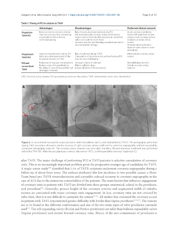
Figure 2. A concomitant myocardial revascularization and transcatheter aortic valve implantation (TAVI). The aortography performed
during TAVI procedure showed a double stenosis of right coronary artery confirmed by selective angiography and not revealed by
computed tomography scan (A). The coronary artery stenosis was very tight; therefore, the percutaneous treatment was performed
before the TAVI (B). After the percutaneous coronary intervention (PCI), a self-expandable valve was implanted (C).
after TAVI. The major challenge of performing PCI in TAVI patients is selective cannulation of coronary
ostia. This is an increasingly important problem given the progressive younger age of candidates for TAVI.
[50]
A single-center study identified that 5.3% of TAVR recipients underwent coronary angiography during a
follow-up of about three years. The authors attributed this low incidence to two possible causes: a Heart
Team-based pre-TAVR revascularization and a possible reduced recourse to coronary angiography in the
case of ACS due to the numerous comorbidities of the patients. The main factors that influence engagement
of coronary ostia in patients with TAVI are divided into three groups: anatomical, related to the prosthesis,
[51]
and procedural . Generally, greater height of the coronary arteries and augmented width of valsalva
sinuses are associated with easier coronary ostia engagement. In fact, coronary ostia are not covered by
valve skirt, thus it is not difficult to cannulate the ostium [51,52] . All studies that evaluated the coronary access
in patients with TAVI demonstrated greater difficulty with Evolut than Sapien prostheses [51,53,54] . The reasons
are to be found in the different conformation and size of the two main types of valve prostheses currently
[51]
used . The self-expanding valves (Evolut and Portico prostheses) are taller than balloon-expandable valves
(Sapien prostheses) and extend beyond coronary ostia. Hence, if the neo-commissure of prosthesis is