
Page 22 - Read Online
P. 22
Page 4 of 19 Khokhar et al. Mini-invasive Surg 2022;6:2 https://dx.doi.org/10.20517/2574-1225.2021.97
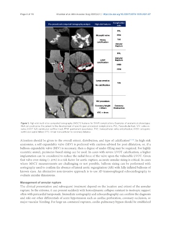
Figure 1. High-risk multi-slice computed tomography (MSCT) features for TAVR complications. Examples of anatomical phenotypes
that can predispose the patient to the development of specific peri-procedural complications. PVL: Paravalvular leak; ViV: valve-in-
valve; LVOT: left ventricular outflow tract; PPM: permanent pacemaker; TVE: transcatheter valve embolization; iVSD: iatrogenic
ventricular septal defect; VTC: virtual transcatheter-to-coronary distance.
Attention should be given to the overall extent, distribution, and type of calcification [37,38] . In high-risk
anatomies, a self-expandable valve (SEV) is preferred with caution advised for post-dilatation, or, if a
balloon-expandable valve (BEV) is necessary, then a degree of under-filling may be required. For highly
eccentric annuli, perimeter-based sizing can be used. In cases with severe LVOT calcification, a higher
implantation can be considered to reduce the radial force of the valve upon the vulnerable LVOT. Given
that valve over-sizing (> 20%) is a risk factor for aortic rupture, accurate annular sizing is critical. In cases
where MSCT measurements are challenging or not possible, balloon sizing can be performed with
aortography used to confirm the absence of lateral aortic regurgitation (AR) with fully inflated balloons of
known sizes. An alternative non-invasive approach is to use 3D transesophageal echocardiography to
evaluate annular dimensions.
Management of annular rupture
The clinical presentation and subsequent treatment depend on the location and extent of the annular
rupture. In the extreme, it can present suddenly with hemodynamic collapse resistant to inotropic support
often with pericardial tamponade. Immediate aortography and echocardiography can confirm the diagnosis
and rule out other differentials of acute hypotension such as cardiac perforation, coronary occlusion, or
major vascular bleeding. For large un-contained ruptures, cardio-pulmonary bypass should be established