
Page 40 - Read Online
P. 40
Chandrasekar et al. Mini-invasive Surg 2021;5:33 https://dx.doi.org/10.20517/2574-1225.2021.12 Page 3 of 16
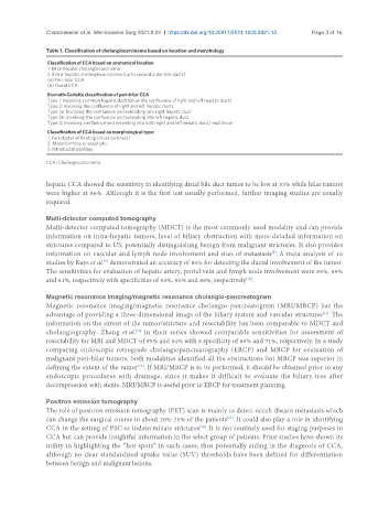
Table 1. Classification of cholangiocarcinoma based on location and morphology
Classification of CCA based on anatomical location
1. Intra-hepatic cholangiocarcinoma
2. Extra-hepatic cholangiocarcinoma (up to second order bile ducts)
(a) Peri-hilar CCA
(b) Distal CCA
Bismuth-Corlette classification of peri-hilar CCA
Type 1: Involving common hepatic duct below the confluence of right and left hepatic ducts
Type 2: Involving the confluence of right and left hepatic ducts
Type 3a: Involving the confluence and extending into right hepatic duct
Type 3b: Involving the confluence and extending into left hepatic duct
Type 4: Involving confluence and extending into both right and left hepatic duct/ multifocal
Classification of CCA based on morphological type:
1. Peri-ductal infiltrating (most common)
2. Mass-forming or exophytic
3. Intraductal papillary
CCA: Cholangiocarcinoma
hepatic CCA showed the sensitivity in identifying distal bile duct tumor to be low at 33% while hilar tumors
were higher at 86%. Although it is the first test usually performed, further imaging studies are usually
required.
Multi-detector computed tomography
Multi-detector computed tomography (MDCT) is the most commonly used modality and can provide
information on intra-hepatic tumors, level of biliary obstruction with more detailed information on
strictures compared to US, potentially distinguishing benign from malignant strictures. It also provides
[8]
information on vascular and lymph node involvement and sites of metastasis . A meta-analysis of 16
studies by Ruys et al. demonstrated an accuracy of 86% for detecting the ductal involvement of the tumor.
[9]
The sensitivities for evaluation of hepatic artery, portal vein and lymph node involvement were 83%, 89%
and 61%, respectively with specificities of 93%, 92% and 88%, respectively .
[10]
Magnetic resonance imaging/magnetic resonance cholangio-pancreatogram
Magnetic resonance imaging/magnetic resonance cholangio-pancreatogram (MRI/MRCP) has the
advantage of providing a three-dimensional image of the biliary system and vascular structures . The
[11]
information on the extent of the tumor/stricture and resectability has been comparable to MDCT and
cholangiography. Zhang et al. in their series showed comparable sensitivities for assessment of
[10]
resectability for MRI and MDCT of 95% and 94% with a specificity of 69% and 71%, respectively. In a study
comparing endoscopic retrograde cholangiopancreatography (ERCP) and MRCP for evaluation of
malignant peri-hilar tumors, both modalities identified all the obstructions but MRCP was superior in
defining the extent of the tumor . If MRI/MRCP is to be performed, it should be obtained prior to any
[12]
endoscopic procedures with drainage, since it makes it difficult to evaluate the biliary tree after
decompression with stents. MRI/MRCP is useful prior to ERCP for treatment planning.
Positron emission tomography
The role of positron emission tomography (PET) scan is mainly to detect occult distant metastasis which
[13]
can change the surgical course in about 20%-25% of the patients . It could also play a role in identifying
[14]
CCA in the setting of PSC or indeterminate strictures . It is not routinely used for staging purposes in
CCA but can provide insightful information in the select group of patients. Prior studies have shown its
utility in highlighting the “hot spots” in such cases, thus potentially aiding in the diagnosis of CCA,
although no clear standardized uptake value (SUV) thresholds have been defined for differentiation
between benign and malignant lesions.