
Page 10 - Read Online
P. 10
Kobayashi et al. Mini-invasive Surg 2020;4:30 I http://dx.doi.org/10.20517/2574-1225.2020.12 Page 5 of 8
Table 2. Causes of postoperative recurrent laryngeal nerve paralysis
Stretch/Traction 5
Compression 3
Thermal/Heat injury 1
Ligature 0
Transection 0
Others (compression in the neck) 1
Table 3. Results of IONM
Evaluation with IONM
+ -
Motion of vocal cord (POD7)
+ 8 2
Pseudo negative
- 2 45
Pseudo positive
+ means loss of motion of vocal cord checked by ENT doctors or loss of response on IONM; - means no signs of paralysis checked by
ENT doctors or adequate response on IONM. IONM: intra-operative neural monitoring; POD: postoperative day; ENT: ear-nose-throat.
Sensitivity: 8/10 = 80%; specificity: 45/47 = 95.7%; positive predictive values: 8/10 = 80%; negative predictive values: 45/47 = 95.7%
A B C
D E F
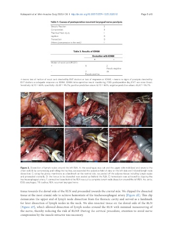
Figure 2. Dissection of lymph nodes around the left RLN. A: the esophagus was cut and the upper side mobilized and sewn to the
chest wall; B: by compressing and rolling the trachea, we expanded the operative field of view on the left side and initiated lymph node
dissection; C: using the glossy membrane as a landmark on the ventral side, we peeled off the adipose tissue including lymph nodes
and proceeded cranially; D: the tissue to be dissected was peeled up behind the RLN; E: hemostasis was achieved by clipping the
tracheoesophageal artery; F: connective tissue behind the RLN was cut to complete lymph node dissection around the left RLN. Ao: aorta;
ESO: esophagus; TR: trachea; RLN: recurrent laryngeal nerve
tissue towards the dorsal side of the RLN and proceeded towards the cranial side. We clipped the dissected
tissue at the most cranial side to achieve hemostasis of the tracheoesophageal artery [Figure 2E]. This clip
demarcates the upper end of lymph node dissection from the thoracic cavity and served as a landmark
for later dissection of lymph nodes in the neck. We also resected tissue on the dorsal side of the RLN
[Figure 2F], which allowed dissection of lymph nodes around the RLN with minimal maneuvering of
the nerve, thereby reducing the risk of RLNP. During the cervical procedure, attention to avoid nerve
compression by the muscle retractor was necessary.