
Page 131 - Read Online
P. 131
Gharagozloo et al. Mini-invasive Surg 2020;4:48 I http://dx.doi.org/10.20517/2574-1225.2020.35 Page 9 of 19
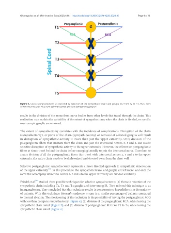
Figure 4. Classic ganglianectomy as depicted by resection of the sympathetic chain and ganglia (X) from T2 to T4. RCA: rami
communicantes albi; RCG: rami communicantes grisei; G: sympathetic ganglion
results in the division of the axons from nerve bodies from other levels that travel through the chain. This
realization may explain the variability of the extent of sympathectomy when the chain is divided, or specific
macroscopic ganglia are removed.
The extent of sympathectomy correlates with the incidence of complications. Disruption of the chain
(sympathectomy), or parts of the chain (sympathicotomy) or removal of selected ganglia will result
in disruption of sympathetic activity to more than just the upper extremity. Only division of the
postganglionic fibers that emanate from the chain and join the intercostal nerves, 2, 3 and 4, can assure
selective disruption of sympathetic activity to the upper extremity. However, the efferent or postganglionic
fibers at times travel behind the chain before emerging laterally to join the intercostal nerve. Therefore, to
assure division of all the postganglionic fibers that travel with intercostal nerves 2, 3 and 4 to the upper
extremity, the entire chain needs to be skeletonized and elevated away from the chest wall.
Selective postganglionic sympathectomy represents a more directed approach to sympathetic denervation
[67]
of the upper extremity . In this procedure, the sympathetic trunk and ganglia are left intact and only the
rami that accompany intercostal nerves 2, 3 and 4 to the upper extremity are divided selectively.
[26]
Friedel et al. studied three possible techniques for selective sympathectomy: (1) thoracic resection of the
sympathetic chain including T2, T3 and T4 ganglia and intervening IR. They referred this technique to as
interganglionare. They concluded that this technique results in compensatory hyperhidrosis in the majority
of patients. With this technique, Horner’s syndrome is seen in a smaller percentage of patients compared
to thermal ablation. The shortcoming of this technique is the possibility of leaving the postganglionic RCG
with less than complete sympathectomy [Figure 4]; (2) division of the preganglionic RCA, while leaving the
sympathetic chain intact [Figure 5]; and (3) division of postganglionic RCG for T2 to T4, while leaving the
sympathetic chain intact [Figure 6].