
Page 36 - Read Online
P. 36
Ardila-Gatas et al. Mini-invasive Surg 2020;4:16 I http://dx.doi.org/10.20517/2574-1225.2019.69 Page 5 of 8
A B C
[6]
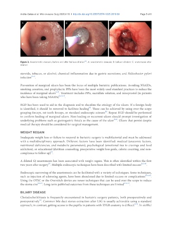
Figure 3. Anastomotic stenosis, before and after balloon dilation . A: anastomotic stenosis; B: balloon dilation; C: anatomosis after
dilation
steroids, tobacco, or alcohol; chemical inflammation due to gastric secretions; and Helicobacter pylori
[2-6]
infection .
Prevention of marginal ulcers has been the focus of multiple bariatric publications. Avoiding NSAIDs,
smoking cessation, and prophylactic PPIs have been the most widely used standard practices to reduce the
[2,3]
incidence of marginal ulcers . Treatment includes PPIs, sucralfate solution, and misoprostol (in patients
who have been taking NSAIDs) [2,4,5,7] .
EGD has been used to aid in the diagnosis and to elucidate the etiology of the ulcers. If a foreign body
[5]
is identified, it should be removed to facilitate healing . These can be achieved by using over the scope
grasping forceps, rat-tooth forceps, or standard endoscopic scissors . Repeat EGD should be performed
[2]
to confirm healing of marginal ulcers. Non-healing or recurrent ulcers should prompt investigation of
[5,6]
underlying problems such as gastrogastric fistula as the cause of the ulcer . Ulcers that persist despite
medical therapy should be considered for surgical management.
WEIGHT REGAIN
Inadequate weight loss or failure to respond to bariatric surgery is multifactorial and must be addressed
with a multidisciplinary approach. Different factors have been identified: medical (anatomic factors,
nutritional deficiencies, and metabolic parameters), psychological (emotional ties to cravings and food
addiction), or educational (dietitian counseling, preoperative weight loss goals, calorie counting, and non-
[7]
compliance to follow up) .
A dilated GJ anastomosis has been associated with weight regain. This is often identified within the first
[7]
two years after surgery . Multiple endoscopic techniques have been described with limited success [2,7,16] .
Endoscopic narrowing of the anastomosis can be facilitated with a variety of techniques. Some techniques,
such as injection of sclerosing agents, have been abandoned due to limited success or complications [2-4,16] .
Using the OTSC or the Overstitch device are newer techniques that can be used over the scope to reduce
the stoma size [16,17] . Long-term published outcomes from these techniques are limited [2-6,17] .
BILIARY DISEASE
Choledocholithiasis is frequently encountered in bariatric surgery patients, both preoperatively and
postoperatively . Common bile duct stones extraction after LSG is usually achievable using a standard
[2]
[2-4]
approach; in contrast, getting access to the papilla in patients with RYGB anatomy is difficult . In skillful