
Page 35 - Read Online
P. 35
Page 4 of 8 Ardila-Gatas et al. Mini-invasive Surg 2020;4:16 I http://dx.doi.org/10.20517/2574-1225.2019.69
A B
[5]
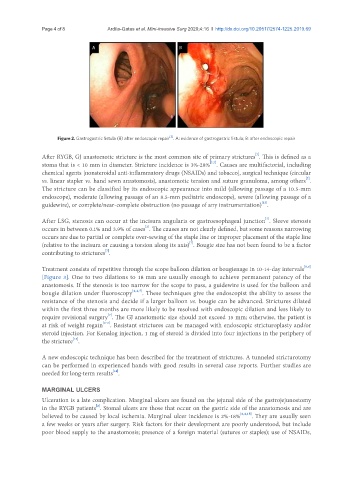
Figure 2. Gastrogastric fistula (B) after endoscopic repair . A: evidence of gastrogastric fistula; B: after endoscopic repair
[7]
After RYGB, GJ anastomotic stricture is the most common site of primary strictures . This is defined as a
[2,7]
stoma that is < 10 mm in diameter. Stricture incidence is 3%-28% . Causes are multifactorial, including
chemical agents [nonsteroidal anti-inflammatory drugs (NSAIDs) and tobacco], surgical technique (circular
[7]
vs. linear stapler vs. hand sewn anastomosis), anastomotic tension and suture granuloma, among others .
The stricture can be classified by its endoscopic appearance into mild (allowing passage of a 10.5-mm
endoscope), moderate (allowing passage of an 8.5-mm pediatric endoscope), severe (allowing passage of a
[12]
guidewire), or complete/near-complete obstruction (no passage of any instrumentation) .
[7]
After LSG, stenosis can occur at the incisura angularis or gastroesophageal junction . Sleeve stenosis
[2]
occurs in between 0.1% and 3.9% of cases . The causes are not clearly defined, but some reasons narrowing
occurs are due to partial or complete over-sewing of the staple line or improper placement of the staple line
[7]
(relative to the incisura or causing a torsion along its axis) . Bougie size has not been found to be a factor
[7]
contributing to strictures .
[5,6]
Treatment consists of repetitive through the scope balloon dilation or bougienage in 10-14-day intervals
[Figure 3]. One to two dilations to 18 mm are usually enough to achieve permanent patency of the
anastomosis. If the stenosis is too narrow for the scope to pass, a guidewire is used for the balloon and
bougie dilation under fluoroscopy [2,4,7] . These techniques give the endoscopist the ability to assess the
resistance of the stenosis and decide if a larger balloon vs. bougie can be advanced. Strictures dilated
within the first three months are more likely to be resolved with endoscopic dilation and less likely to
[7]
require revisional surgery . The GJ anastomotic size should not exceed 15 mm; otherwise, the patient is
[2-6]
at risk of weight regain . Resistant strictures can be managed with endoscopic stricturoplasty and/or
steroid injection. For Kenalog injection, 1 mg of steroid is divided into four injections in the periphery of
[13]
the stricture .
A new endoscopic technique has been described for the treatment of strictures. A tunneled stricturotomy
can be performed in experienced hands with good results in several case reports. Further studies are
[14]
needed for long-term results .
MARGINAL ULCERS
Ulceration is a late complication. Marginal ulcers are found on the jejunal side of the gastrojejunostomy
[2]
in the RYGB patients . Stomal ulcers are those that occur on the gastric side of the anastomosis and are
believed to be caused by local ischemia. Marginal ulcer incidence is 2%-18% [2,4,15] . They are usually seen
a few weeks or years after surgery. Risk factors for their development are poorly understood, but include
poor blood supply to the anastomosis; presence of a foreign material (sutures or staples); use of NSAIDs,