
Page 34 - Read Online
P. 34
Ardila-Gatas et al. Mini-invasive Surg 2020;4:16 I http://dx.doi.org/10.20517/2574-1225.2019.69 Page 3 of 8
A B
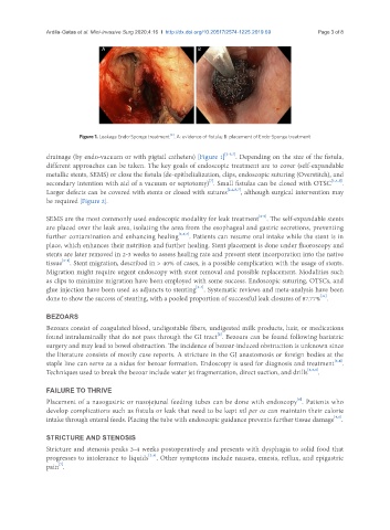
Figure 1. Leakage Endo-Sponge treatment . A: evidence of fistula; B: placement of Endo-Sponge treatment
[6]
drainage (by endo-vacuum or with pigtail catheters) [Figure 1] [2-5,7] . Depending on the size of the fistula,
different approaches can be taken. The key goals of endoscopic treatment are to cover (self-expandable
metallic stents, SEMS) or close the fistula (de-epithelialization, clips, endoscopic suturing (Overstitch), and
[7]
secondary intention with aid of a vacuum or septotomy) . Small fistulas can be closed with OTSC [2,4,5] .
Larger defects can be covered with stents or closed with sutures [2,4,5,7] , although surgical intervention may
be required [Figure 2].
[2-6]
SEMS are the most commonly used endoscopic modality for leak treatment . The self-expandable stents
are placed over the leak area, isolating the area from the esophageal and gastric secretions, preventing
further contamination and enhancing healing [2,6,7] . Patients can resume oral intake while the stent is in
place, which enhances their nutrition and further healing. Stent placement is done under fluoroscopy and
stents are later removed in 2-3 weeks to assess healing rate and prevent stent incorporation into the native
tissue . Stent migration, described in > 40% of cases, is a possible complication with the usage of stents.
[2-4]
Migration might require urgent endoscopy with stent removal and possible replacement. Modalities such
as clips to minimize migration have been employed with some success. Endoscopic suturing, OTSCs, and
[2-4]
glue injection have been used as adjuncts to stenting . Systematic reviews and meta-analysis have been
done to show the success of stenting, with a pooled proportion of successful leak closures of 87.77% .
[11]
BEZOARS
Bezoars consist of coagulated blood, undigestable fibers, undigested milk products, hair, or medications
[2]
found intraluminally that do not pass through the GI tract . Bezoars can be found following bariatric
surgery and may lead to bowel obstruction. The incidence of bezoar-induced obstruction is unknown since
the literature consists of mostly case reports. A stricture in the GJ anastomosis or foreign bodies at the
[2,4]
staple line can serve as a nidus for bezoar formation. Endoscopy is used for diagnosis and treatment .
Techniques used to break the bezoar include water jet fragmentation, direct suction, and drills [2,5,6] .
FAILURE TO THRIVE
[6]
Placement of a nasogastric or nasojejunal feeding tubes can be done with endoscopy . Patients who
develop complications such as fistula or leak that need to be kept nil per os can maintain their calorie
[4,6]
intake through enteral feeds. Placing the tube with endoscopic guidance prevents further tissue damage .
STRICTURE AND STENOSIS
Stricture and stenosis peaks 3-4 weeks postoperatively and presents with dysphagia to solid food that
progresses to intolerance to liquids . Other symptoms include nausea, emesis, reflux, and epigastric
[2,4]
[7]
pain .