
Page 37 - Read Online
P. 37
Page 4 of 11 Abdalla et al. Mini-invasive Surg 2019;3:39 I http://dx.doi.org/10.20517/2574-1225.2019.38
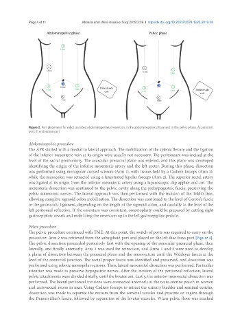
Figure 2. Port placement for robot-assisted abdominoperineal resection, in the abdominopelvic phase and in the pelvic phase. A: assistant
port; E: endoscope port
Abdominopelvic procedure
The APR started with a medial to lateral approach. The mobilization of the splenic flexure and the ligation
of the inferior mesenteric vein at its origin were usually not necessary. The peritoneum was incised at the
level of the sacral promontory. The avascular presacral plane was entered, and this plane was developed
identifying the origin of the inferior mesenteric artery and the left ureter. During this phase, dissection
was performed using monopolar curved scissors (Arm 1), with tissues held by a Cadiere forceps (Arm 3),
while the mesocolon was retracted using a fenestrated bipolar forceps (Arm 2). The superior rectal artery
was ligated at its origin from the inferior mesenteric artery using a laparoscopic clip applier and cut. The
mesenteric dissection was continued to the pelvic cavity along the prehypogastric fascia, preserving the
pelvic autonomic nerves. The lateral approach was then performed with the incision of the Toldt’s line,
allowing complete sigmoid colon mobilization. The dissection was continued to the level of Gerota’s fascia
or the gastrocolic ligament, depending on the length of the sigmoid colon, and caudally to the level of the
left peritoneal reflection. If the omentum was consistent, omentoplasty could be prepared by cutting right
gastroepiploic vessels and mobilizing the omentum up to the left gastroepiploic pedicle.
Pelvic procedure
The pelvic procedure continued with TME. At this point, the switch of ports was required to carry on the
procedure: Arm 2 was retrieved from the subxiphoid port and placed on the left iliac fossa port [Figure 2].
The pelvic dissection proceeded posteriorly first with the opening of the avascular presacral plane, then
laterally, and finally anteriorly. Arm 3 was used for retraction, and Arms 1 and 2 were used to develop
a plane of dissection between the presacral plane and the mesorectum until the Waldeyer fascia at the
level of the anorectal junction. The rectal proper fascia was identified and preserved, and dissection was
performed using robotic monopolar scissors. Then, lateral mesorectal dissection was performed. Particular
attention was made to preserve hypogastric nerves. After the incision of the peritoneal reflection, lateral
pelvic attachments were divided distally, until the levator ani. Lastly, the anterior mesorectal dissection was
performed. The lateral peritoneal incisions were connected anteriorly at the recto-uterine pouch in women
and rectovesical recess in men. Using Cadiere forceps to retract the urinary bladder and seminal vesicles,
dissection was made to separate the rectum from the seminal vesicles and prostate or vagina through
the Denonvillier’s fascia, followed by separation of the levator muscles. When pelvic floor was reached