
Page 36 - Read Online
P. 36
Abdalla et al. Mini-invasive Surg 2019;3:39 I http://dx.doi.org/10.20517/2574-1225.2019.38 Page 3 of 11
A B
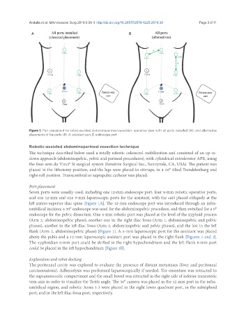
Figure 1. Port placement for robot-assisted abdominoperineal resection: operative view with all ports installed (A); and alternative
placements of the ports (B). A: assistant port; E: endoscope port
Robotic-assisted abdominoperineal resection technique
The technique described below used a totally robotic colorectal mobilization and consisted of an up-to-
down approach (abdominopelvic, pelvic and perineal procedures), with cylindrical extralevator APR, using
the four-arm da Vinci® Si surgical system (Intuitive Surgical Inc., Sunnyvale, CA, USA). The patient was
placed in the lithotomy position, and the legs were placed in stirrups, in a 20° tilted Trendelenburg and
right-roll position. Transurethral or suprapubic catheter was placed.
Port placement
Seven ports were usually used, including one 12-mm endoscope port, four 8-mm robotic operative ports,
and one 12-mm and one 5-mm laparoscopic ports for the assistant, with the cart placed obliquely at the
left antero-superior iliac spine [Figure 1A]. The 12-mm endoscope port was introduced through an infra-
umbilical incision; a 30° endoscope was used for the abdominopelvic procedure, and then switched for a 0°
endoscope for the pelvic dissection. One 8-mm robotic port was placed at the level of the xyphoid process
(Arm 2, abdominopelvic phase), another one in the right iliac fossa (Arm 1, abdominopelvic and pelvic
phases), another in the left iliac fossa (Arm 3, abdominopelvic and pelvic phases), and the last in the left
flank (Arm 2, abdominopelvic phase) [Figure 2]. A 5-mm laparoscopic port for the assistant was placed
above the pubis and a 12-mm laparoscopic assistant port was placed in the right flank [Figures 1 and 2].
The xyphoidian 8-mm port could be shifted in the right hypochondrium and the left flank 8-mm port
could be placed in the left hypochondrium [Figure 1B].
Exploration and robot docking
The peritoneal cavity was explored to evaluate the presence of distant metastases (liver and peritoneal
carcinomatosis). Adhesiolysis was performed laparoscopically if needed. The omentum was retracted to
the supramesocolic compartment and the small bowel was retracted in the right side of inferior mesenteric
vein axis in order to visualize the Treitz angle. The 30° camera was placed in the 12-mm port in the infra-
umbilical region, and robotic Arms 1-3 were placed in the right lower quadrant port, in the subxiphoid
port, and in the left iliac fossa port, respectively.