
Page 102 - Read Online
P. 102
Parthasarathi et al. Mini-invasive Surg 2019;3:20 I http://dx.doi.org/10.20517/2574-1225.2019.10 Page 3 of 14
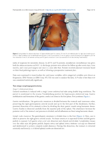
Figure 1. Port position for abdominal phase. A: Supraumbilical port for camera: 10 mm; B: Left midclavicular for right hand working port:
5 mm; C: Right midclavicular for left hand working port: 5 mm (converted to 12 mm for gastric conduit formation); D: Epigastric port for
liver retraction 5 mm; E: Left anterior axillary port for gastric retraction: 5 mm (converted to 12 mm for dividing Transhiatally)
index of suspicion for metastatic disease. In all T3 and T4 patients, neoadjuvant chemotherapy was given
both for adenocarcinoma and SCC. At discharge, patients were advised for follow up after seven days, three
months, and 1-year post-surgery and once in a year after that. Patients received adjuvant treatment based
on their final pathology reports, as well as following a preoperative treatment plan.
Data were expressed in mean/median for continuous variables, while categorical variables were shown as
frequencies. SPSS Version 24 (IBM Corp. NY, US) was used to analyze the data. A P-value of less than 0.05
was considered statistically significant.
Two-stage esophagogastrectomy
Stage I: Abdominal phase
General anesthesia is induced with a single lumen endotracheal tube using double lung ventilation. The
patient is positioned in the reverse Trendelenburg position for laparoscopic abdominal step. Gastric
mobilization and formation of the gastric conduit are done in the first phase. Port positions: Figure 1.
Gastric mobilization: The gastrocolic omentum is divided between the stomach and transverse colon,
preserving the right gastroepiploic arterial arcade and up to the first part of the duodenum. Further,
proximal dissection of the stomach is done by dividing the short gastric vessels using harmonic shears.
Gastric fundus is dissected carefully from the superior pole of the spleen. The attachment between the
posterior wall of the stomach and pancreas is divided, and all adhesions are freed using harmonic shears.
Lymph node clearance: The gastrohepatic omentum is divided close to the liver [Figure 2]. Here, care is
taken to preserve the right gastric arterial arcade. The lesser omentum is separated further and left gastric
pedicle is exposed. Left gastric artery and vein dissected and clipped and divided. Lymphofatty tissues
over the celiac axis, left gastric pedicle, hepatic artery and splenic artery (stations 7,8,9,11p) are dissected
and skeletonized to achieve complete nodal clearance. During this dissection, the stomach is retracted
anteriorly and laterally. 11 d (distal splenic) nodes are dissected out.