
Page 103 - Read Online
P. 103
Page 4 of 14 Parthasarathi et al. Mini-invasive Surg 2019;3:20 I http://dx.doi.org/10.20517/2574-1225.2019.10
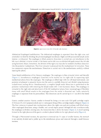
Figure 2. Laparoscopic view after D2 lymph node clearance
Abdominal Esophageal mobilization: The abdominal esophagus is separated from the right crus, and
anteriorly it is freed by dividing the phrenoesophageal membrane. Right and left paracardial lymph nodes
(station 1,2) dissected. The esophagus is lifted, posterior dissection is carried out, and attachment to the
left crus is divided. Anterior border of the hiatus and/or left crus are divided obliquely toward the left side
using harmonic shears. Sometimes, the division of the right crus may also be needed to facilitate dissection
into the posterior mediastinum. Then liver retractor is advanced into the mediastinum for retraction. These
two maneuvers expose the mediastinum. Dissection is carried out in the mediastinum carefully without
injuring the pleura.
Trans hiatal mobilization of the thoracic esophagus: The esophagus is then retracted above and laterally
[Figure 3]. Intrathoracic esophageal dissection to be started on the right side by separating right
mediastinal pleura from the esophagus. The esophagus is lifted high with the left-hand instrument, and
posterior attachment to preaortic fascia over the aorta is carefully dissected and divided using harmonic
scalpel/vessel sealing device. Simultaneously, a complete lymph nodal dissection is performed in the
posterior mediastinum with removing the lymph nodes with 5 mm harmonic shears. The esophagus is
retracted to the right side and attachment of the left mediastinal pleura from mesoesophagus (Fibrofatty
tissue with small blood vessels to the esophagus from the aorta) is dissected out. Lastly, anteriorly
esophagus is separated from the pericardium. This dissection can be carried up to 6-7 cm above the hiatal
level.
Gastric conduit creation: Gastric conduit is formed by firing a 60 mm endo GIA gold cartridge stapler
(© Ethicon US, LLC) perpendicularly and 2-3 subsequent firing of blue cartridge staplers obliquely [Figure 4].
The liver retractor is passed into mediastinum above the right crus and pericardium well lifted above.
After esophageal dissection, using a flexible, and curved stapler passed through left side intrathoracically,
the esophagus is divided, making sure, the transection line is above the tumor [Figure 5]. A small opening
formed in the right pleura and the gastric conduit is placed within in the right pleural cavity [Figure 6].
Through a Pfannenstiel incision, the specimen is removed out. In cases of smaller lesions, the stomach
is not entirely divided and is pulled up in the intrathoracic phase and removed through a small thoracic
incision.