
Page 28 - Read Online
P. 28
Page 6 of 11 Cicero et al. Mini-invasive Surg 2019;3:25 I http://dx.doi.org/10.20517/2574-1225.2018.012
A B
C D
C
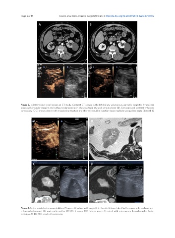
Figure 7. Indeterminate renal lesions at CT study. Contrast CT shows in the left kidney voluminous, partially exophitic, hypodense
lesion with irregular margins and without enhancement in phases arterial (A) and venous phase (B). Greyscale and contrast-enhanced
sonography (C-D) shows a lesion with mixed echo structure and after microbubble injection shows multiple vascularized septa (Bosniak 4)
A B
C D
Figure 8. Fusion-guided microwave ablation. 75 years old patient with exophitic in the right kidney, identified by sonography and contrast-
enhanced ultrasound (A) and confirmed by MR (B), it was a RCC (biopsy proven) treated with microwaves through guided fusion
technique (C-D). RCC: renal cell carcinoma