
Page 25 - Read Online
P. 25
Cicero et al. Mini-invasive Surg 2019;3:25 I http://dx.doi.org/10.20517/2574-1225.2018.012 Page 3 of 11
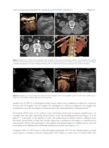
A B
C D
Figure 1. Pseudotumor. A: Grey-scale sonography shows isoechoic cortical mass (or dromedary hump?) in the midportion of the kidney
(arrows); on contrast-enhanced sonograms the lesion shows enhancement equal (or similar) to the kidney in all vascular phases.
Computed tomography with contrast injection before (B), after (C) and 3D reconstruction (D) doesn’t show focal renal lesions
A B
Figure 2. Bosniak 2 cyst. A: Grey scale and contrast enhanced sonograms shows small exophitic hypoechoic mass with smooth margin in
lower pole; B: Completely avascular after microbubble injection
Another use of CEUS is to distinguish kidney tumors and/or other conditions in which the convective
B mode and US Doppler have not helpful the radiologist to a definitive diagnosis: for example, the
pseudotumors have the same degree of enhancement as the remaining healthy renal parenchyma .
[7,8]
Particularly CEUS seems to be useful in some mimicking anatomical variations: pseudotumors, for
example, have the same enhancing characteristics of the surrounding parenchyma [Figure 1] in all
phases [9,10] . Conversely, in the majority of cases, the enhancement in kidney tumors is different from
the nearly parenchyma: at least one vascular phase has a variation in the degree or distribution of
enhancement. CEUS is also appropriate in the analysis and characterization of complex kidney cysts and
particularly in the differentiation of Bosniak classification of renal cysts [Figures 2-5].
Compared with CT, CEUS seems to have the higher preciseness of CT for the characterization of renal
cystic lesions according to lesional enhancement (LE). Infact, in some cases, CT and/or RMI can’t