
Page 155 - Read Online
P. 155
Westwood et al. Mini-invasive Surg 2018;2:38 I http://dx.doi.org/10.20517/2574-1225.2018.50 Page 7 of 11
Table 2. Three-point grading system for the assessment of the plane of anal canal/sphincter dissection in abdominoperineal
excision specimens for low-rectal cancer
Grade Description
Extra-levator plane The specimen has a cylindrical shape due to the presence of levator ani removed en bloc with the mesorectum and
sphincters. Any defects must be no deeper than 5 mm. No waisting of the specimen. Smooth CRM on slicing
Sphincteric plane The specimen is waisted and the CRM in this region is formed by the surface of the sphincter muscles which have
been removed intact
Intrasphincteric plane The specimen is waisted and includes deviations into the sphincter muscles, submucosa and complete perforations
CRM: circumferential resection margin
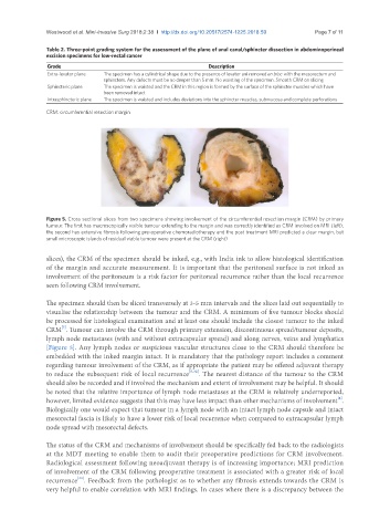
Figure 5. Cross sectional slices from two specimens showing involvement of the circumferential resection margin (CRM) by primary
tumour. The first has macroscopically visible tumour extending to the margin and was correctly identified as CRM involved on MRI (left);
the second has extensive fibrosis following pre-operative chemoradiotherapy and the post treatment MRI predicted a clear margin, but
small microscopic islands of residual viable tumour were present at the CRM (right)
slices), the CRM of the specimen should be inked, e.g., with India ink to allow histological identification
of the margin and accurate measurement. It is important that the peritoneal surface is not inked as
involvement of the peritoneum is a risk factor for peritoneal recurrence rather than the local recurrence
seen following CRM involvement.
The specimen should then be sliced transversely at 3-5 mm intervals and the slices laid out sequentially to
visualise the relationship between the tumour and the CRM. A minimum of five tumour blocks should
be processed for histological examination and at least one should include the closest tumour to the inked
[5]
CRM . Tumour can involve the CRM through primary extension, discontinuous spread/tumour deposits,
lymph node metastases (with and without extracapsular spread) and along nerves, veins and lymphatics
[Figure 5]. Any lymph nodes or suspicious vascular structures close to the CRM should therefore be
embedded with the inked margin intact. It is mandatory that the pathology report includes a comment
regarding tumour involvement of the CRM, as if appropriate the patient may be offered adjuvant therapy
to reduce the subsequent risk of local recurrence [5,32] . The nearest distance of the tumour to the CRM
should also be recorded and if involved the mechanism and extent of involvement may be helpful. It should
be noted that the relative importance of lymph node metastases at the CRM is relatively underreported,
[4]
however, limited evidence suggests that this may have less impact than other mechanisms of involvement .
Biologically one would expect that tumour in a lymph node with an intact lymph node capsule and intact
mesorectal fascia is likely to have a lower risk of local recurrence when compared to extracapsular lymph
node spread with mesorectal defects.
The status of the CRM and mechanisms of involvement should be specifically fed back to the radiologists
at the MDT meeting to enable them to audit their preoperative predictions for CRM involvement.
Radiological assessment following neoadjuvant therapy is of increasing importance; MRI prediction
of involvement of the CRM following preoperative treatment is associated with a greater risk of local
recurrence . Feedback from the pathologist as to whether any fibrosis extends towards the CRM is
[33]
very helpful to enable correlation with MRI findings. In cases where there is a discrepancy between the