
Page 153 - Read Online
P. 153
Westwood et al. Mini-invasive Surg 2018;2:38 I http://dx.doi.org/10.20517/2574-1225.2018.50 Page 5 of 11
Table 1. Three-point grading system for the assessment of the plane of mesorectal dissection in total mesorectal excision/
anterior resection specimens for rectal cancer
Grade of excision Quality of surgery Description
Mesorectal Good surgery Intact smooth mesorectal surface with only minor irregularities. Any defects must be no
deeper than 5 mm. No coning of the specimen distally. Smooth CRM on slicing
Intramesorectal Moderate surgery Moderate bulk to mesorectum but irregularity of the mesorectal surface. Moderate distal
coning. Muscularis propria not visible with the exception of levator insertion. Moderate
irregularity of CRM on slicing
Muscularis propria Poor surgery Little bulk to mesorectum with defects down onto the muscularis propria and/or very
irregular CRM. It includes perforations through the CRM
CRM: circumferential resection margin
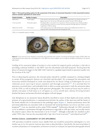
Figure 2. Cross sectional slices showing a mesorectal plane specimen with smooth circumferential resection margin (CRM) (left) and
intramesorectal plane specimen with irregularity to the CRM and obvious defects, but no evidence of defects extending to the muscle
tube (right)
Grading of the mesorectal plane of excision is a key marker for surgical quality and plays a vital role in
providing continual feedback to the MDT team for educational and audit purposes. Feeding back the
planes of mesorectal surgery in the MRC CR07 trial led to a gradual improvement in specimen quality over
[26]
the duration of the trial .
Prior to dissecting the specimen, the external surface should be carefully examined by a histopathologist
to ensure all key prognostic features are identified and described. We recommend the description and
dissection method developed in Leeds and adopted by the Royal College of Pathologists for use in
colorectal cancer reporting to ensure consistent and thorough assessment [5,27] . It is essential that the
specimen is received intact, and preferably fresh, to allow accurate assessment of both the mesorectal plane
and the CRM, as well as taking the whole specimen photographs. The mesorectal fascia may be easier to
identify and assess in fresh tissue as it will appear as a shiny smooth layer, whereas following formalin
fixation the fascia can become distorted and appear dull and opaque.
Any disruptions in the mesorectal fascia should be described in terms of their depth and extent, and the
presence of surgical perforation, a communication between the surface of the specimen and the lumen of
the bowel, should also be documented in the pathology report [Figure 3]. Tumour perforations above the
peritoneal reflections are associated with an increased risk of intraperitoneal recurrence and have a poor
[29]
[28]
prognosis ; these are classified as pT4a using TNM8 staging . Perforations through the CRM commonly
occur in addition to perforations through the peritoneum, especially in APE specimens, and usually involve
[19]
the anterior aspect, where the mesorectum is at its thinnest . Although technically not classified as pT4a
[20]
[30]
under TNM rules, these are also associated with a high risk of local recurrence and reduced survival .
PATHOLOGICAL ASSESSMENT OF APE SPECIMENS
APE specimens should be received intact, ideally fresh and should be assessed by pathologists in a very
similar way to that for anterior resection specimens as described above. Specifically, the mesorectal plane of
excision and presence of intraoperative perforations should be evaluated and specimen photographs should