
Page 154 - Read Online
P. 154
Page 6 of 11 Westwood et al. Mini-invasive Surg 2018;2:38 I http://dx.doi.org/10.20517/2574-1225.2018.50
Figure 3. Anterior resection specimen showing a large anterior perforation with a defect into the lumen of the bowel (left), which is
confirmed on cross sectional slicing where part of the anterior rectal wall is missing (right)
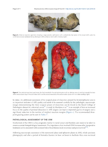
Figure 4. Two abdominoperineal specimens, the first resected in the sphincteric plane with an obvious area of waisting towards the base
of the mesorectum/top of the sphincters (left) and the second resected in the extra-levator plane with no visible waisting (right)
be taken. An additional assessment of the surgical plane of dissection around the levator/sphincter area is
an important indicator of APE quality and needs to be assessed carefully by the pathologist; macroscopic
images demonstrating the three surgical planes of dissection can be found in the Royal College of
[31]
[5]
Pathologists dataset for colorectal cancer . A study by Martijnse et al. is an example of how an increased
focus on the quality of perineal dissection in APE surgery can lead to improved quality of surgery and a
significant reduction in the number of involved resection margins [Figure 4]. The recommended three-
point grading system can be seen in Table 2 .
[3]
PATHOLOGICAL ASSESSMENT OF THE CRM
Involvement of the CRM is a key prognostic marker in rectal cancer and therefore care needs to be taken to
ensure accurate histopathological assessment. The importance of an involved CRM increases after preoperative
treatment as it is associated with increased risk of local failure, local recurrence and poor survival [9,18] .
Following macroscopic assessment of the mesorectal plane (and sphincter planes in APE), whole specimen
photography and after a period of formalin fixation (at least 48 hours to facilitate thin cross sectional