
Page 17 - Read Online
P. 17
Page 4 of 13 Lesch et al. Mini-invasive Surg 2023;7:25 https://dx.doi.org/10.20517/2574-1225.2023.31
Table 1. Variable influences on an abdominal wall load
Peak Plateau Continuous
Low level High level
Mode of load Cough Dynamic lift IPAP/ICU Valsalva
[1]
Amount of impacts 425 Few Continuous Once
Impact area Normal/perpendicular strain variable Shear strain
Tensile load Youngs elastic modulus Poisson’s ratio Shear modulus
Interrelated and not to be derived from each other due to anisotropy
Materials to be tested any any any
Impact area Small or large Small or large large
Conditions Wet Wet Wet
Temperature Room or body Room or body Room or body
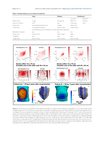
Figure 2. (A) Analysis of assessment of the abdominal walls of a patient with computed tomography (CT) at rest and during the
Valsalva maneuver (CTAV) in two herniated patient cases (left and right picture group). The panel shows the shift and the distortion in
red shades. The area of greatest movement is shown in bright red, fading with the amount of movement. (B) CT scans of the abdomen
during the Valsalva maneuver of non-herniated patients. Right: a female patient with a weight gain of 30 kg presented with mild pain two
years after reconstruction with a slight distension on the left-hand side and a circumscript detection of protrusion on the right-hand side
by analysis with artificial intelligence (right middle picture) with a shift of the ventral abdominal wall of maximally 16 mm (far right
picture) Left: a male patient with a weight gain of 25 kg presenting with pain after playing soccer two years after the reconstruction (L-
shaped incision for liver transplantation) with a bulge in the right lateral extension of the L-shaped incision without a reherniation (far
left picture) and a shift of the ventral abdominal wall of 41 mm as a maximum (left middle picture).