
Page 15 - Read Online
P. 15
Nishimura Percutaneous endoscopic cervical laminectomy
Table 1: Summary of the detailed features of the 11 cases with PECL
Case Age, Original Affected Approach Operation Hospital Follow-up mJOA mJOA Recovary
No. years Gender disease level side time, min stay, periods, Complication score score rate, %
months
days
pre-op post-op
1 59 Male HYL C5/6 Left 96 5 28 No 11 15 66.7
2 61 Male HYL + Disc C4/5 Left 81 5 25 No 10.5 13 36.5
3 67 Male OPLL C5/6, 6/7 Right 141 5 24 No 11 15 66.7
4 72 Female HYL + Disc C4/5 Right 85 8 20 No 10 13 42.9
5 58 Male HYL C3/4 Left 70 3 17 No 11.5 16 81.8
6 65 Female HYL C3/4 Right 74 5 17 No 12 15 60.0
7 71 Female OPLL C4/5 Right 75 8 13 No 11.5 16 81.8
8 76 Male HYL + Disc C5/6 Right 82 8 12 No 11 13 33.3
9 69 Male HYL C5/6 Left 95 8 10 No 10 13 42.9
10 52 Male OPLL C6/7 Left 88 3 10 No 11 15 66.7
11 59 Female HYL C5/6 Left 71 5 7 No 10 13 42.9
PECL: percutaneous endoscopic cervical laminectomy; mJOA: modified Japanese Orthopaedic Association; HYL: hypertrophic yellow
ligament; Disc: cervical disc protrusion; OPLL: oscificated posterior longitiduinal ligament
Data analysis the ethical standards of the committee and with the
Pre- and postoperative neurological statuses were Helsinki Declaration.
evaluated using the modified Japanese Orthopaedic
Association (mJOA) score for cervical myelopathy. RESULTS
Recovery rate was calculated as follows: recovery rate
= postoperative mJOA - preoperative mJOA/17 (full Eleven patients were registered for this study. The
score) - preoperative mJOA score × 100. [10,11] Statistical mean age was 64.5 years (range 52-76 years) and the
analysis was performed using Student’s t-test. P values male/female ratio was 7/4. The most affected vertebral
less than 0.05 were considered statistically significant. level was C5/6 (5 cases), followed by C4/5 (3 cases).
Detailed information on each case, such as operation
This retrospective study was approved by the ethics time, follow-up period, and hospital stay, is shown in
committee of the Wakayama Koyo Hospital, and Table 1.
informed consent was obtained from the patients
for publication of this study and any accompanying During the mean follow-up period of 16.6 months (range
images. The procedures were in accordance with 7-28 months), the mJOA score improved significantly
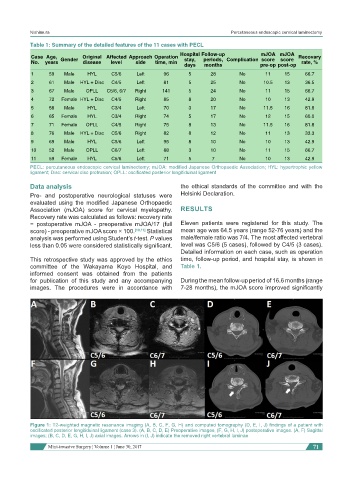
Figure 1: T2-weighted magnetic resonance imaging (A, B, C, F, G, H) and computed tomography (D, E, I, J) findings of a patient with
oscificated posterior longitiduinal ligament (case 3). (A, B, C, D, E) Preoperative images; (F, G, H, I, J) postoperative images. (A, F) Sagittal
images; (B, C, D, E, G, H, I, J) axial images. Arrows in (I, J) indicate the removed right vertebral laminae
Mini-invasive Surgery ¦ Volume 1 ¦ June 30, 2017 71