
Page 34 - Read Online
P. 34
Magaribuchi et al. Mini-invasive Surg 2024;8:6 https://dx.doi.org/10.20517/2574-1225.2023.81 Page 3 of 8
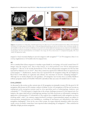
Figure 1. (A) Generation of a 3D kidney model through 3D reconstruction from CT scans; (B) 3D printed model, which is fabricated from
the kidney’s 3D model using a 3D printer, aids in understanding anatomical features such as the kidney, tumor, and blood vessels; (C)
3D navigation via VR. The 3D model of the kidney can be viewed from various angles using devices such as a head-mounted display; (D)
3D navigation via AR. By overlaying the 3D kidney model onto the actual surgical view, detailed anatomical features can be grasped
during surgery. No direct patient identifiers are included in this image. 3D: Three-dimensional; CT: computed tomography; VR: virtual
reality; AR: augmented reality.
surgical or head-mounted displays to provide surgeons with navigation [22-24] . In VR navigation, there is no
overlay (registration) of 3D models onto the surgical view.
AR
AR is a method that allows surgeons to visualize organ details by overlaying a 3D model created from CT
images onto the surgical view. Due to this feature, it is often preferred over VR for intraoperative
navigation . The use of 3D models derived from CT for AR in intraoperative navigation for PN was first
[13]
reported by Ukimura et al. in 2008 . Since then, numerous reports have followed about its application in
[25]
actual surgery [26-32] . Furthermore, studies have also reported that 3D navigation using AR can display the
[33]
blood flow of the kidney in segments and enhance the outcomes of selective clamping strategies .
Although not as widely adopted as echo guidance, 3D navigation has become more accessible thanks to
software programs (such as SYNAPSE VINCENT and others) that can create 3D models from CT scans.
CHALLENGES
As discussed in the section on the current state of 3D navigation in minimally invasive PN, the proof of 3D
navigation effectiveness in PN remains a subject of debate. In fact, 3D navigation in PN has not become as
widespread as the navigation systems used in other specialties such as otolaryngology, dentistry, and
orthopedics [34,35] . This discrepancy is largely due to the unique challenges presented by the surgical area. For
instance, the organs dealt with in otolaryngology undergo very little movement or deformation, making the
application of preoperative CT information to the surgical site straightforward, with navigation accuracy
reaching under 1 mm . In contrast, organs involved in urology or gastrointestinal surgery are subject to
[36]
intraoperative manipulations and insufflation, resulting in movement and deformation that make
navigation challenging . Even in the case of the rectum, an organ relatively immobile within the pelvic
[37]
[38]
cavity, errors of around 19mm have been reported when attempting 3D navigation . This confirms the
difficulty of high-precision navigation.