
Page 12 - Read Online
P. 12
Page 6 of 21 Tsuboi et al. Mini-invasive Surg 2024;8:26 https://dx.doi.org/10.20517/2574-1225.2023.94
TM
TM
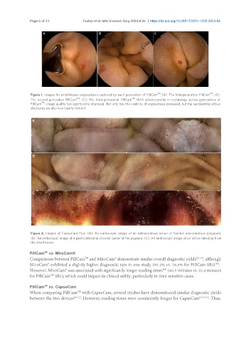
Figure 1. Images for small-bowel angioectasia captured by each generation of PillCam . (A): The first-generation PillCam ; (B):
TM
TM
The second-generation PillCam ; (C): The third-generation PillCam . With advancements in technology across generations of
TM
PillCam , image quality has significantly improved. Not only has the visibility of angioectasia increased, but the surrounding villous
structures are also now clearly visible.X.
Figure 2. Images of CapsoCam Plus. (A): An endoscopic image of an adenomatous lesion of familial adenomatous polyposis;
(B): An endoscopic image of a gastrointestinal stromal tumor of the jejunum; (C): An endoscopic image of an active bleeding from
the small bowel.
PillCam vs. MiroCam®
TM
Comparisons between PillCam and MiroCam® demonstrate similar overall diagnostic yields [66-68] , although
TM
MiroCam® exhibited a slightly higher diagnostic rate in one study (95.2% vs. 78.6% for PillCam SB2) .
[68]
However, MiroCam® was associated with significantly longer reading times (40.3 minutes vs. 25.4 minutes
[68]
for PillCam SB2), which could impact its clinical utility, particularly in time-sensitive cases.
TM
TM
PillCam vs. CapsoCam
When comparing PillCam with CapsoCam, several studies have demonstrated similar diagnostic yields
TM
between the two devices [69-72] . However, reading times were consistently longer for CapsoCam [69,70,72] . Thus,