
Page 71 - Read Online
P. 71
Neumann et al. J Transl Genet Genom 2022;6:353-60 https://dx.doi.org/10.20517/jtgg.2022.06 Page 355
CASE REPORT
Our patient was a 27-year-old woman who presented initially with a thyroid mass to an outside hospital,
which was diagnosed by biopsy as squamous cell carcinoma. The patient presented to our hospital for
further management. She underwent a partial excisional biopsy of the thyroid mass, with final pathology
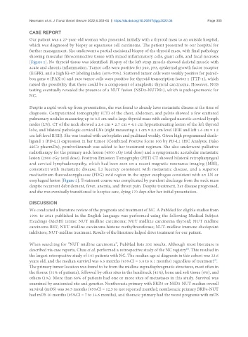
showing muscular fibroconnective tissue with mixed inflammatory cells, giant cells, and focal necrosis
[Figure 1]. No thyroid tissue was identified. Biopsy of the left strap muscle showed skeletal muscle with
acute and chronic inflammation. Tumor cells were positive for p40, p53, epidermal growth factor receptor
(EGFR), and a high Ki-67 labeling index (40%-70%). Scattered tumor cells were weakly positive for paired-
box gene 8 (PAX-8) and rare tumor cells were positive for thyroid transcription factor 1 (TTF-1), which
raised the possibility that there could be a component of anaplastic thyroid carcinoma. However, NGS
testing eventually revealed the presence of a NUT fusion (NSD3-NUTM1), which is pathognomonic for
NC.
Despite a rapid work-up from presentation, she was found to already have metastatic disease at the time of
diagnosis. Computerized tomography (CT) of the chest, abdomen, and pelvis showed a few scattered
pulmonary nodules measuring up to 0.5 cm and a large thyroid mass with enlarged necrotic cervical lymph
nodes (LN). CT of the neck showed a 2.6 cm × 2.7 cm × 4.1 cm hypoattenuating lesion of the left thyroid
lobe, and bilateral pathologic cervical LNs (right measuring 3.1 cm × 2.2 cm level II/III and left 1.6 cm × 1.2
cm left level II/III). She was treated with carboplatin and paclitaxel weekly. Given high programmed death-
ligand 1 (PD-L1) expression in her tumor (Combined Positive Score 100 by PD-L1 IHC Analysis, Dako
22C3 pharmDx), pembrolizumab was added to her treatment regimen. She also underwent palliative
radiotherapy for the primary neck lesion (4500 cGy total dose) and a symptomatic acetabular metastatic
lesion (2000 cGy total dose). Positron Emission Tomography (PET) CT showed bilateral retropharyngeal
and cervical lymphadenopathy, which had been seen on a recent magnetic resonance imaging (MRI),
consistent with metastatic disease, L2 lucency consistent with metastatic disease, and a superior
mediastinum fluorodeoxyglucose (FDG) avid region in the upper esophagus consistent with an LN or
esophageal lesion [Figure 2]. Treatment course was complicated by purulent discharge from the neck mass
despite recurrent debridement, fever, anemia, and throat pain. Despite treatment, her disease progressed,
and she was eventually transitioned to hospice care, dying 170 days after her initial presentation.
DISCUSSION
We conducted a literature review of the prognosis and treatment of NC. A PubMed for eligible studies from
1991 to 2021 published in the English language was performed using the following Medical Subject
Headings (MeSH) terms: NUT midline carcinoma; NUT midline carcinoma thyroid; NUT midline
carcinoma BET; NUT-midline carcinoma histone methyltransferase; NUT-midline immune checkpoint
inhibitors; NUT-midline treatment. Results of the literature helped drive treatment for our patient.
When searching for “NUT midline carcinoma”, PubMed lists 202 results. Although most literature is
described via case reports, Chau et al. performed a retrospective study of the NC registry . This resulted in
[8]
the largest retrospective study of 141 patients with NC. The median age at diagnosis in this cohort was 23.6
years old, and the median survival was 6.5 months (95%CI = 5.8 to 9.1 months) regardless of treatment .
[7]
The primary tumor location was found to be from the midline supradiaphragmatic structures, most often in
the thorax (51% of patients), followed by other sites in the head/neck (41%), bone and soft tissue (6%), and
others (1%). More than 60% of patients had one or more sites of metastases in this study. Survival was
examined by anatomical site and genetics. Nonthoracic primary with BRD3 or NSD3-NUT median overall
survival (mOS) was 36.5 months (95%CI = 12.5 to not reported months); nonthoracic primary BRD4-NUT
had mOS 10 months (95%CI = 7 to 14.6 months), and thoracic primary had the worst prognosis with mOS