
Page 72 - Read Online
P. 72
Page 356 Neumann et al. J Transl Genet Genom 2022;6:353-60 https://dx.doi.org/10.20517/jtgg.2022.06
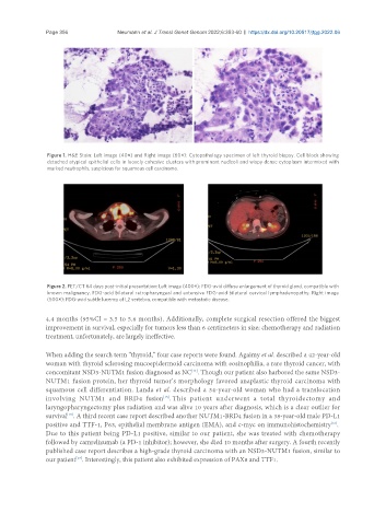
Figure 1. H&E Stain: Left image (40×) and Right image (60×): Cytopathology specimen of left thyroid biopsy. Cell block showing
detached atypical epithelial cells in loosely cohesive clusters with prominent nucleoli and wispy dense cytoplasm intermixed with
marked neutrophils, suspicious for squamous cell carcinoma.
Figure 2. PET/CT 64 days post-initial presentation: Left image (400×): FDG-avid diffuse enlargement of thyroid gland, compatible with
known malignancy. FDG-acid bilateral retropharyngeal and extensive FDG-avid bilateral cervical lymphadenopathy. Right image
(500×): FDG-avid subtle lucency of L2 vertebra, compatible with metastatic disease.
4.4 months (95%CI = 3.5 to 5.6 months). Additionally, complete surgical resection offered the biggest
improvement in survival, especially for tumors less than 6 centimeters in size; chemotherapy and radiation
treatment, unfortunately, are largely ineffective.
When adding the search term “thyroid,” four case reports were found. Agaimy et al. described a 42-year-old
woman with thyroid sclerosing mucoepidermoid carcinoma with eosinophilia, a rare thyroid cancer, with
concomitant NSD3-NUTM1 fusion diagnosed as NC . Though our patient also harbored the same NSD3-
[14]
NUTM1 fusion protein, her thyroid tumor’s morphology favored anaplastic thyroid carcinoma with
squamous cell differentiation. Landa et al. described a 34-year-old woman who had a translocation
involving NUTM1 and BRD4 fusion . This patient underwent a total thyroidectomy and
[15]
laryngopharyngectomy plus radiation and was alive 10 years after diagnosis, which is a clear outlier for
survival . A third recent case report described another NUTM1-BRD4 fusion in a 38-year-old male PD-L1
[15]
positive and TTF-1, P63, epithelial membrane antigen (EMA), and c-myc on immunohistochemistry .
[16]
Due to this patient being PD-L1 positive, similar to our patient, she was treated with chemotherapy
followed by camrelizumab (a PD-1 inhibitor); however, she died 10 months after surgery. A fourth recently
published case report describes a high-grade thyroid carcinoma with an NSD3-NUTM1 fusion, similar to
our patient . Interestingly, this patient also exhibited expression of PAX8 and TTF1.
[17]