
Page 54 - Read Online
P. 54
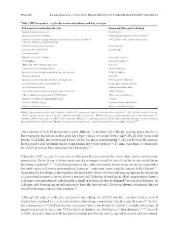
Page 338           Estévez-Arias et al. J Transl Genet Genom 2022;6:333-52  https://dx.doi.org/10.20517/jtgg.2022.04

               Table 1. CMT therapeutics: target cell processes and pathways and drug strategies
                Cell process or mechanism of action                      Compound/therapeutic strategy
                Reduction of gene expression                             Ascorbic acid
                Reduction of protein synthesis                           Progesterone antagonists and modulators
                Inhibition of Schwann cells proliferation and reduction of protein synthesis   PXT3003 (baclofen, sorbitol, naltrexone)
                (baclofen: GABA receptor modulator)
                          B
                Partial silencing of gene expression                     Gene silencing
                Gene insertion (AAV1-NT3)                                Gene therapy
                Gene substitution
                Regulation of myelin thickness                           Neuregulin pathways
                UPR inhibition                                           Curcumin, sephin-1
                TRPA1 and TRPV1 channels activation                      FLX-787
                Prevention of axonal degeneration                        SARM1 inhibitors
                Reduction of microtubules acetylation (axonal transport)  HDAC6 inhibitors
                Myostatin pathway                                        ACE-083
                Reduction of abnormal calcium influx in Schwann cells    P2X7 receptor modulators
                Correction of defective lipid biosynthesis               Dietary lipid supplementation
                Nav 1.8 channel blocking                                 Sodium channel blockers
                Decreased number/activity of macrophages in the nerve    CSF1R inhibitors
                PIKfyve inhibition and decrease of PI3,5P2 levels        PIKfyve enzyme inhibitors
                Reduction of neurotoxic deoxysphingolipids               L-Serine
                Purine nucleotides supply                                S-adenosylmethionine (SAM)
                Inhibition of aldose reductase                           Aldose reductase inhibitors
               GABA : Gamma-aminobutyric acid B receptor; AAV1-NT3: adeno-associated virus-mediated neurotrophin-3; UPR: unfolded protein response;
                   B
               TRPA1: transient receptor potential cation channel, subfamily A, member 1; TRPV1: transient receptor potential cation channel subfamily V
               member; SARM1: sterile alpha and TIR motif-containing 1; HDAC6: histone deacetylase 6; CSF1R: colony-stimulating factor 1 receptor; PIKfyve:
               phosphatidylinositol 3-phosphate 5-kinase. Summarized information in this table was extracted from Pisciotta et al. (2021) [50] .


               The scenario of GDAP1 mutations is quite different from other CMT disease-causing genes due to its
               heterogeneity: mutations in this gene have been related to axonal forms (AR-CMT2K with vocal cord
               paresis (CMT2K), an intermediate form (CMTRIA), and a demyelinating (CMT4A) form of the disease.
               Both recessive and dominant modes of inheritance have been reported . To date, more than 100 mutations
                                                                          [56]
               in GDAP1 gene have been related to CMT phenotype .
                                                            [59]
               Clinically, CMT caused by mutations in this gene is characterized by severe distal motor and sensory
               neuropathy. Nevertheless, a diverse spectrum of phenotypes should be considered due to the variability in
               inheritance patterns [56,60,61] . It has been proposed that, while autosomal recessive mutations are responsible
               for early-onset and severe neuropathies, dominant mutations cause a milder course of the disease .
                                                                                                       [62]
               Regarding the histological abnormalities, the sural nerve biopsy of severe affected segregating the disease in
               an autosomal recessive manner shows a pronounced depletion of myelinated fibers, regenerative clusters
               and signs of axonal atrophy. Additionally, a small proportion of thin myelinated fibers and proliferation of
               Schwann cells forming onion bulb structures have also been found. The most relevant cytoplasmic feature
                                                  [63]
               would be the mitochondrial abnormalities .
               Although the explicit molecular mechanism underlying the GDAP1 function remains unclear, several
                                                                                                 [59]
               studies have explored its role in mitochondria physiology: morphology, function, and dynamics . Firstly,
               the consequence of GDAP1 mutations can impair these mitochondrial functions through mitochondrial
               membrane potential reduction, ATP production changes, or a disbalance of their dynamics [57,64,65] . Second,
               GDAP1 may also interact with transport proteins involved in mitochondrial transport and movement.