
Page 42 - Read Online
P. 42
Casas-Alba et al. J Transl Genet Genom 2022;6:322-32 https://dx.doi.org/10.20517/jtgg.2022.03 Page 326
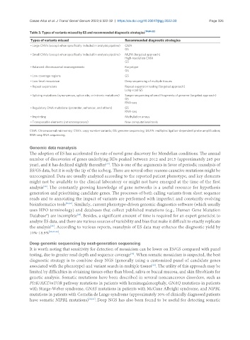
Table 3. Types of variants missed by ES and recommended diagnostic strategies [10,20-22]
Types of variants missed Recommended diagnostic strategies
• Large CNVs (except when specifically included in analysis pipeline) CMA
GS
• Small CNVs (except when specifically included in analysis pipeline) MLPA (targeted approach)
High-resolution CMA
GS
• Balanced chromosomal rearrangements Karyotype
GS
• Low coverage regions GS
• Low-level mosaicism Deep sequencing of multiple tissues
• Repeat expansions Repeat expansion testing (targeted approach)
Long-read GS
• Splicing mutations (synonymous, splice site, or intronic mutations) Sanger sequencing of small fragments of genome (targeted approach)
GS
RNA-seq
• Regulatory DNA mutations (promoter, enhancer, and others) GS
RNA-seq
• Imprinting Methylation arrays
• Transposable elements (retrotransposons) New computational tools
CMA: Chromosomal microarray; CNVs: copy number variants; GS: genome sequencing; MLPA: multiplex ligation-dependent probe amplification;
RNA-seq: RNA sequencing.
Genomic data reanalysis
The adoption of ES has accelerated the rate of novel gene discovery for Mendelian conditions. The annual
number of discoveries of genes underlying RDs peaked between 2012 and 2015 (approximately 285 per
year), and it has declined slightly thereafter . This is one of the arguments in favor of periodic reanalysis of
[24]
ES/GS data, but it is only the tip of the iceberg. There are several other reasons causative mutations might be
unrecognized. Data are usually analyzed according to the reported patient phenotype, and key elements
might not be available to the clinical laboratory or might not have emerged at the time of the first
analysis . The constantly growing knowledge of gene networks is a useful resource for hypothesis
[25]
generation and prioritizing candidate genes. The processes of both calling variants from short sequence
reads and to annotating the impact of variants are performed with imperfect and constantly evolving
bioinformatics tools [25,26] . Similarly, current phenotype-driven genomic diagnostics software (which usually
uses HPO terminology) and databases that collect published mutations (e.g., Human Gene Mutation
[25]
Database®) are incomplete . Besides, a significant amount of time is required for an expert geneticist to
analyze ES data, and there are various sources of variability and bias that make it difficult to exactly replicate
[25]
the analysis . According to various reports, reanalysis of ES data may enhance the diagnostic yield by
10%-18.9% [25,27-29] .
Deep genomic sequencing by next-generation sequencing
It is worth noting that sensitivity for detection of mosaicism can be lower on ES/GS compared with panel
[20]
testing, due to greater read depth and sequence coverage . When somatic mosaicism is suspected, the best
diagnostic strategy is to combine deep NGS (generally using a customized panel of candidate genes
associated with the phenotype) and variant search in multiple tissues . The utility of this approach may be
[30]
limited by difficulties in obtaining tissues other than blood, saliva or buccal mucosa, and skin fibroblasts for
genetic analysis. Somatic mutations have been described in several noncancerous disorders, such as
PI3K/AKT/mTOR pathway mutations in patients with hemimegalencephaly, GNAQ mutations in patients
with Sturge-Weber syndrome, GNAS mutations in patients with McCune Albright syndrome, and NIPBL
mutations in patients with Cornelia de Lange syndrome (approximately 30% of clinically diagnosed patients
have somatic NIPBL mutations) [30,31] . Deep NGS has also been found to be useful for detecting somatic