
Page 17 - Read Online
P. 17
Jafri et al. J Transl Genet Genom 2022;6:281-9 https://dx.doi.org/10.20517/jtgg.2021.63 Page 283
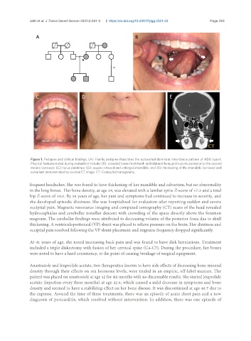
Figure 1. Pedigree and clinical findings. (A): Family pedigree illustrates the autosomal dominant inheritance pattern of ADO type I.
Physical features noted during evaluation include; (B): crowded lower front teeth and bilateral bony protrusions posterior to the second
molars (arrows); (C): torus palatinus; (D): square-shaped and enlarged mandible; and (E): thickening of the mandible (arrows) and
calvarium demonstrated by coronal CT image. CT: Computed tomography.
frequent headaches. She was found to have thickening of her mandible and calvarium, but no abnormality
in the long bones. Her bone density, at age 34, was elevated with a lumbar spine Z-score of +7.3 and a total
hip Z-score of +8.0. By 36 years of age, her pain and symptoms had continued to increase in severity, and
she developed episodic dizziness. She was hospitalized for evaluation after reporting sudden and severe
occipital pain. Magnetic resonance imaging and computed tomography (CT) scans of the head revealed
hydrocephalus and cerebellar tonsillar descent with crowding of the space directly above the foramen
magnum. The cerebellar findings were attributed to decreasing volume of the posterior fossa due to skull
thickening. A ventriculoperitoneal (VP) shunt was placed to relieve pressure on the brain. Her dizziness and
occipital pain resolved following the VP shunt placement and migraine frequency dropped significantly.
At 41 years of age, she noted increasing back pain and was found to have disk herniations. Treatment
included a triple diskectomy with fusion of her cervical spine (C4-C7). During the procedure, her bones
were noted to have a hard consistency, to the point of causing breakage of surgical equipment.
Anastrazole and leuprolide acetate, two therapeutics known to have side effects of decreasing bone mineral
density through their effects on sex hormone levels, were trialed in an empiric, off-label manner. The
patient was placed on anastrozole at age 42 for six months with no discernable results. She started leuprolide
acetate (injection every three months) at age 42.6, which caused a mild decrease in symptoms and bone
density and seemed to have a stabilizing effect on her bone disease. It was discontinued at age 46.7 due to
the expense. Around the time of these treatments, there was an episode of acute chest pain and a new
diagnosis of pericarditis, which resolved without intervention. In addition, there was one episode of