
Page 95 - Read Online
P. 95
Ma et al. J Transl Genet Genom 2022;6:179-203 https://dx.doi.org/10.20517/jtgg.2021.48 Page 193
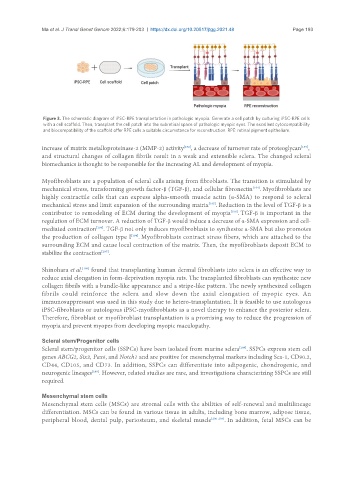
Figure 3. The schematic diagram of iPSC-RPE transplantation in pathologic myopia. Generate a cell patch by culturing iPSC-RPE cells
with a cell scaffold. Then, transplant the cell patch into the subretinal space of pathologic myopic eyes. The excellent cytocompatibility
and biocompatibility of the scaffold offer RPE cells a suitable circumstance for reconstruction. RPE: retinal pigment epithelium.
[192]
[193]
increase of matrix metalloproteinase-2 (MMP-2) activity , a decrease of turnover rate of proteoglycan ,
and structural changes of collagen fibrils result in a weak and extensible sclera. The changed scleral
biomechanics is thought to be responsible for the increasing AL and development of myopia.
Myofibroblasts are a population of scleral cells arising from fibroblasts. The transition is stimulated by
mechanical stress, transforming growth factor-β (TGF-β), and cellular fibronectin . Myofibroblasts are
[194]
highly contractile cells that can express alpha-smooth muscle actin (α-SMA) to respond to scleral
mechanical stress and limit expansion of the surrounding matrix . Reduction in the level of TGF-β is a
[195]
contributor to remodeling of ECM during the development of myopia . TGF-β is important in the
[196]
regulation of ECM turnover. A reduction of TGF-β would induce a decrease of α-SMA expression and cell-
mediated contraction . TGF-β not only induces myofibroblasts to synthesize α-SMA but also promotes
[196]
the production of collagen type I . Myofibroblasts contract stress fibers, which are attached to the
[194]
surrounding ECM and cause local contraction of the matrix. Then, the myofibroblasts deposit ECM to
stabilize the contraction .
[197]
Shinohara et al. found that transplanting human dermal fibroblasts into sclera is an effective way to
[198]
reduce axial elongation in form-deprivation myopia rats. The transplanted fibroblasts can synthesize new
collagen fibrils with a bundle-like appearance and a stripe-like pattern. The newly synthesized collagen
fibrils could reinforce the sclera and slow down the axial elongation of myopic eyes. An
immunosuppressant was used in this study due to hetero-transplantation. It is feasible to use autologous
iPSC-fibroblasts or autologous iPSC-myofibroblasts as a novel therapy to enhance the posterior sclera.
Therefore, fibroblast or myofibroblast transplantation is a promising way to reduce the progression of
myopia and prevent myopes from developing myopic maculopathy.
Scleral stem/Progenitor cells
[199]
Scleral stem/progenitor cells (SSPCs) have been isolated from murine sclera . SSPCs express stem cell
genes ABCG2, Six2, Pax6, and Notch1 and are positive for mesenchymal markers including Sca-1, CD90.2,
CD44, CD105, and CD73. In addition, SSPCs can differentiate into adipogenic, chondrogenic, and
neurogenic lineages . However, related studies are rare, and investigations characterizing SSPCs are still
[199]
required.
Mesenchymal stem cells
Mesenchymal stem cells (MSCs) are stromal cells with the abilities of self-renewal and multilineage
differentiation. MSCs can be found in various tissue in adults, including bone marrow, adipose tissue,
peripheral blood, dental pulp, periosteum, and skeletal muscle [200-204] . In addition, fetal MSCs can be